登录


Wollastonite from Poudrette quarry, Mont Saint-Hilaire, La Vallée-du-Richelieu RCM, Montérégie, Québec, Canada
K8G-PF0Wollastonite Ca3(Si3O9)

Field of View: 3.9 mm
Compact mass of coarse wollastonite fibers.
Photographed by and in the collection of Douglas Merson.
Obtained from László Horváth
753-N8GWollastonite Ca3(Si3O9)

Field of View: 4.3 mm
This massive white wollastonite was obtained from Bob Rothenberg.
1WK-TQTWollastonite Ca3(Si3O9)


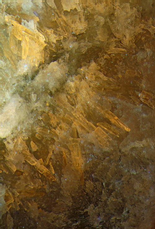
Field of View: 4.7 mm
Found 8/16/97.
This is a 3-way comparison of SW & MW UV fluorescence and visible light images using the Mindat interactive comparison tool. The MW/SW comparison isn’t “apples to apples”. Using 5W LED flashlights positioned ca 6 cm form the specimen, with ISO set to 800 on my camera, and objective magnification 2x on my scope, each stacked frame for the SW image required an exposure of 15 sec. Each MW frame required only 1 sec.
Compared to pectolite, wollastonite is considered to be a rather uncommon mineral at MSH. However, since getting my new LED SW & MW LED flashlights in 2022, I am finding that it isn’t really all that uncommon in the linings of marble xenolith cavities. But well-formed, or even isolated crystals, do seem to be rare. This specimen exhibits some of the best crystals that I have spotted so far. I will be so bold as to claim that a few of them may even be decently terminated.
Under visible light, it is almost impossible to distinguish the wollastonite in xenolith linings from very similar looking pectolite in the same environment. (Crystals of pectolite in the cavities themselves are another matter. But wollastonite rarely occurs in the cavities.) As shown in these images, even under MW UV, the “salmon” response of wollastonite looks much like that of typical pectolite at MSH. But the “gold” SW response is very distinctive. (According to the most recently published list of MSH fluorescent species – Horváths et al, Can Min SP 14, 2019, Appendix 6 – pectolite can also fluoresce weak to moderate orange under SW, MW and LW UV. But all of the SW “orange” fluorescing stuff from marble xenoliths that I have - not counting organic “cavity scum” - always fluoresces “salmon” – not orange – under MW. I.e. it is wollastonite. I have yet to find any orange fluorescing pectolite from MSH.)
The background of the wollastonite crystals, which fluoresces somewhat like some pectolite is apparently actually mostly massive calcite. It fizzes vigorously in HCl.
This material is part of the cavity lining of a marble xenolith specimen. The cavity lining consists mostly of fluorescent pectolite and wollastonite, the latter being more abundant. There is also some weakly fluorescent massive calcite. The cavity lining also has some very brightly fluorescent cuspidine. See [https://www.mindat.org/photo-1329281html].)
The cavity itself (not shown) has both fluorescent and non-fluorescent pectolite crystals, very dark hibschite, a large “convoluted” crystal of annite, and small, but nice, fluorapatite crystals. The latter fluoresce almost the same colors as cuspidine, but have typical prismatic fluorapatite habits. (See [https://www.mindat.org/photo-1307494.html].)
Note: The mages under the last two links will also show up as “related” if you click on this image and look in the TN list below it.)
W2L-JFDWollastonite Ca3(Si3O9) , Cuspidine Ca8(Si2O7)2F4
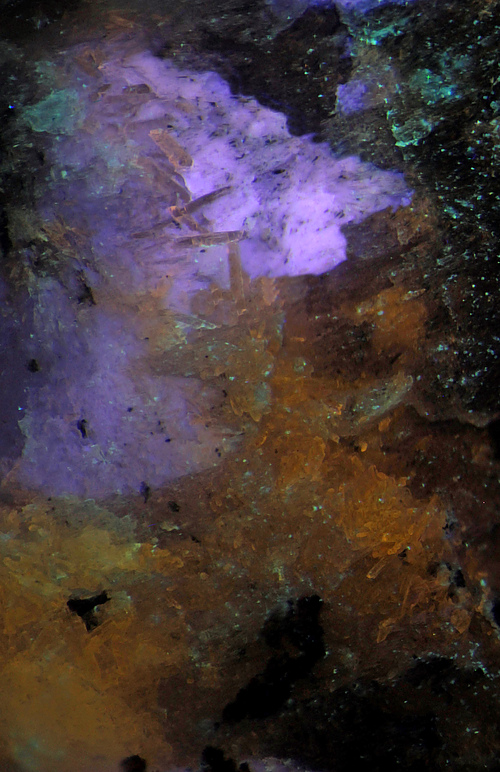
Field of View: 4.5 mm
Found 8/16/97. Analyzed find.
This is a 3-way comparison of SW & MW UV fluorescence and visible light images using the Mindat interactive comparison too. The SW/MW comparison isn’t “apples to apples” because, as as usual with the 5W LED flashlights that I use, the MW response is actually stronger. With the flashlights positioned ca 6 cm from the specimen, and objective magnification 2X, each (stacked) frame for the SW image required an exposure of 10 sec with ISO set to 800 on my camera. Each MW frame required only 1/3 sec using ISO 400. That is really, really bright as fluorescent responses go. If this were a larger specimen it would, as they say, “blow your socks off” under MW.
I have posted several photos of cuspidine (and wollastonite) from MSH in the past year or so (2023). The MW response of the cuspidine is more or less consistently bright blue. The SW response varies, from almost nothing to pale blue. The response on this specimen is pinker than most. Cuspidine at MSH almost always comes in characteristically “spear-head” shaped crystal. In this case, you have to use your imagination to see diffuse spear-heads. Nonetheless, I think it is still cuspidine because of the UV responses, the association with wollastonite in a marble xenolith cavity lining, and because of the lack of a better alternative ID. (It isn't pectolite because pectolite at MSH almost never glows under SW.) For more on the cuspidine (and wollastonite) ID see [https://www.mindat.org/photo-1297994.html], under which you will find EDS and XRD data – and more blather. (This specimen is from the same find.)
My main reason for posting yet another set of cuspidine/wollastonite images is that this is the clearest example of the inclusion of well-formed wollastonite crystals in cuspidine. In fact, it is also the clearest example of any wollastonite crystals at MSH that I have. (The wollastonite is characteristically “gold” under SW UV and “salmon” under MW UV. Both colors look more like their namesakes to the naked-eye than depicted here.) On some other specimens, cuspidine exhibits inclusions that fluoresce greenish, but I have not been able to locate one that can be photographed too well as of yet.
As is typical for these two species at MSH, spotting them under visible light is “mission impossible” – which probably explains why cuspidine was not recognized until recently. The specimen was actually collected for its platy apophyllite-(K), but on this specimen, that does not fluoresce under either SW or MW. The specimen also has some small dark “phlogopite” crystals, probably annite at least in part., and an unusual dark amphibole (unidentified). These last two are in a cavity that is out of view in these images.
I have already mentioned that the MW response of the cuspidine is very bright (and the wollastonite is no slouch either). To quantify this a bit further, the response looks about as bright as depicted with the MW flashlight as far as 1 m or more from the specimen – even with some lights on. To see an equally bright SW response, the SW flashlight has to be within 10-12 cm of the specimen.
N2N-21RWollastonite Ca3(Si3O9) , Calcite CaCO3 , Sphalerite ZnS

Field of View: 4.7 mm
Found Oct 1994.
This is a comparison of MW & SW UV fluorescence and visible light images. With the 5 W LED flashlights that I use, the SW response is actually much weaker than the MW response. The SW image required an exposure 10 times as long as the MW image. See below for details.
In the MW image, the pinkish glow is from wollastonite. To my naked eye, the color looks more “salmon”, which is one of the MW responses for MSH wollastonite listed in Appendix 6 of Can Min SP 14 (2019), but sometimes my camera knows better so I didn’t try to adjust the recorded color too much. This response is very similar to that of much of the pectolite from MSH, but the “gold” SW response is characteristic of MSH wollastonite. (According to SP 14, MSH pectolite can also have a weak orange SW response, but in that case it has a moderate MW response. In any case, I have never seen such responses from MSH pectolite.)
The blue glow in the MW image is from calcite. Blue responses are unusual but not rare for MSH calcite. In any case, the stuff fizzes in HCl, so it isn’t fluorite. See [https://www.mindat.org/photo-1364938.html] for a close-up MW vs. visible light comparison.
I don’t know what the pale yellow spots are. I also don’t know what the stuff (upper right hand corner) that glows orange under both MW and SW is. (Note: This stuff is out of focus because it is actually on the back of the specimen – i.e. far from the area that I stacked. A closer look reveals that it too is mostly wollastonite but it glows fairly orange-yellow on some edges and other patches. I don't know if these areas are intrinsic to the wollastonite or just some sort of crust. But the ornage-yellow is brighter than the "salmon" of the wollastonite so - even there is not a great deal of it, it "takes over" in an out of focus image. The only mystery is what this crust - or whatever - is.)
The sphalerite is the dark crystal seen in all three images. See [https://www.mindat.org/photo-313276.html] for a stereo visible light view which also shows the calcite more clearly
With the LED flashlights positioned ca 5-6 cm from the specimen and using objective magnification 2X on my scope with ISO set to 800 on my camera, each frame for the (stacked) SW image required an exposure of 25 sec. Each MW frame required only 2.5 sec. Hand –held in a semi-darkened room, the MW response looks about as bright as depicted with the flashlight held as far as 25-30 cm from the specimen. To see the SW response as brightly, the flashlight has to be within ca 5-6 cm of the specimen (and there can’t be too much light in the room).
0YX-GWVWollastonite Ca3(Si3O9) , Miserite K1.5-x(Ca,Y,REE)5(Si6O15)(Si2O7)(OH,F)2·yH2O , Calcite CaCO3

Dimensions: 4.6 cm x 3.5 cm x 0.5 cm
Ex Ron Waddell (via Ewald Gerstmann).
This is a comparison of MW and SW UV fluorescence images using the Mindat interactive comparison tool. (To see both images, click on the TN. You can then use the controls to choose the view you prefer.)
It is also yet another new parent image, posted to show the pinkish MW response, which, with the 5W LED “flashlights” that I now use, is much brighter than the more familiar “gold” SW response. To see the SW response as bright as depicted, a 5W, 255 nm, flashlight needs to be ca 8-10 cm from the specimen. The MW response is about equally bright with a 310 nm flashlight held ca 40-50 cm from the specimen. (However, note that in the MW image, the top and bottom are poorly lit because I had the flashlight too close to the specimen.)
According to the most recently published list of MSH fluorescent minerals (Horváths et al, Can Min SP 14, 2019, Appendix 6), the MW response of wollastonite is “salmon”. To me – on this specimen at least – it looks pink. The magenta-red areas are calcite. (The calcite does not glow under SW – as is typical at MSH.) The dark prisms are miserite (see the child photos for visible light images).
The SW response is much weaker than that of wollastonite from “Franklin” (NJ). On the other hand Sterling Mine wollastonite (which is all that I have) is essentially “dead” under MW. In fact, judging from previous posts on Mindat, the strong pink MW response shown here appears to be very unusual for the species.
For more details, see the earlier parent photos (all of which have been retained as child photos).
ANJ-LWTCuspidine Ca8(Si2O7)2F4 , Wollastonite Ca3(Si3O9)

Dimensions: 4 cm x 3 cm x 3 cm
Found 8/16/97.
This is a comparison of MW UV fluorescence and visible light images using the Mindat interactive comparison tool. (To see both images, click on the TN. You can then use the controls to choose the view you prefer
In the MW image, the salmon colored glow is from wollastonite. This is the typical MW response of MSH wollastonite per Can Min SP 14 (2019), Appendix 6. The SW response is “gold” – also typical of MSH wollastonite.
The bright blue glow is likely from cuspidine, the ID of which has finally been established via SEM-EDS, Raman, and “pseudo-Gandolfi” XRD thanks to Joy Desor. Of these analyses, the most convincing (to me) are the SEM-EDS and XRD. I have posted all of these results as “analysis” under [https://www.mindat.org/photo-1297996.html], which see for more details. Note: While I am fairly confident about the “cuspidine” ID, I have not yet added it to the MSH species list pending comments from some MSH experts.
May 22 2023: Cuspidine has now been added to the MSH species list.
The red patches at the bottom are from calcite, and the pinkish-violet is from pectolite. The little bit of greenish glow isn’t “dirt”. It is coming from a small, radiating spray, which look like some elpidite. This glows yellow under SW, which is also common for elpidite. One does not expect to see elpidite in a marble xenolith, but it’s not unheard of.
The visible light image looks like a photo of “nothing”. That’s because it is actually a side view of a specimen of platy fluorapophyllite-(K) and pectolite. From what I have seen, cuspidine is found only in the lining of marble xenolith cavities at MSH – i.e. the part that most collectors try to trim away. It is also nearly impossible to spot under visible light, and the UV response is generally (but not always) much weaker under SW UV. (To a large extent, these comments also apply to MSH wollastonite.)
The MW response of the “cuspidine” is very bright. Using a 5 W, 310 nm, LED flashlight, it will look about as bright as depicted with the flashlight as far as 1 m from the specimen. The SW response with a similar 255 nm flashlight is less intense. The flashlight needs to be ca 10-15 cm from the specimen. The color of the SW response is also more pinkish. See the link above for SW images. (That specimen is from the same find as this one - proably from the same boulder.)
EUD-XTXCuspidine Ca8(Si2O7)2F4 , Wollastonite Ca3(Si3O9)

Dimensions: 3 cm x 3 cm x 4 cm
This is a comparison of SW and MW UV fluorescence images using the Mindat interactive comparison tool. (To see both images, click on the TN. You can then use the controls to choose the view you prefer.)
The comparison isn’t “apples to apples”. With the 5 W LED “flashlights” that I use, the SW response doesn’t appear as bright as depicted unless the 255 nm flashlight is held about 5-6 cm from the specimen. The MW response is much brighter, possibly in part because the MW visible light filter is less effective and probably also blocking less of the UV. In any case, the MW response looks about as bright as depicted with the 310 nm flashlight held about 40-50 cm from the specimen. The posted images look roughly equal because different exposure times were used.
In these images, the blue to lilac-blue responses are from cuspidine, the ID of which has finally been established (on a sample from this specimen) via SEM-EDS, Raman, and “pseudo-Gandolfi” XRD thanks to Joy Desor. Of these analyses, the most convincing (to me) are the SEM-EDS and XRD. I have posted all of these results as “analysis” child photos, which see for more details. Note: While I am fairly confident about the “cuspidine” ID, I have not yet added it to the MSH species list pending comments from some MSH experts.
May 22 2023: Cuspidine has now been added to the MSH species list.
Verification of the “cuspidine” ID proved to be very challenging and time consuming because the crystals are always embedded in the lining of marble xenolith cavities and are almost impossible to spot visually. (See the child visible light vs. SW comparison images.) This makes it very difficult to obtain a “pure” sample for analysis. In this case, the cuspidine is embedded mostly in wollastonite, which glows a very characteristic “gold” under SW UV and “salmon” under MW UV. The latter is similar to the pinkish response of most MSH pectolite, but MSH pectolite glows only very weakly if at all under SW UV, so while the wollastonite has not yet been analyzed, I am fairly sure of the ID.
It is somewhat ironic that this specimen finally provided an unambiguous sample for analysis because many of the larger cuspidine crystals on it (such as the one in the center) have inclusions of wollastonite clearly visible under a scope. (See the close-up MW vs. SW vs. comparison of one such crystal.) In other cases, the cuspidine is embedded in pectolite, sometimes with fluorite and even with hackmanite.
Note added June 19, 2024.
When I first started taking the cuspidine ID seriously, I noticed that at least some of the cuspidine (as on this specimen) had a distinct phosphorescence but I didn't have time to document what I was seeing. I have just now taken another look at this specimen and can report the following:
Under MW there is bright red-pink "flash" of short duration. It's not quite as bright as the blue MW response itself, but nearly so.
Under SW there is a weaker violet-pink "flash" of similar duration. Again, the "flash" isn't quite as bright as the SW response itself.
At this point I don't know how typical these responses are, but it should be noted that the SW response itself is brighter than that of most of the other cuspidine specimens from MSH that I have.
I also investigated the phosphorescence of the wollastonite:
Under MW the wollastonite glows bright salmon pink (similar to much of the pectolite from MSH), but the phosphorescent "flash" is bright orange and somewhat longer lasting than the cuspidine "flash".
Under SW the wollastonite glows weaker "gold" and there is a weaker "flash" of a slightly more yellowish color.
These wollastonite responses are about the same as those listed in Can Min SP 14 (2019) Appendix 6, except that I wouldn't call either the MW or SW duration "long". At most, what I see is "slow flash". I do have some other wollastonite specimens from MSH with brighter and slightly longer phosphorescence.
Attempts to photograph the phosphorescence were all dismal failures.
Note added Oct 18 2024: I have finally purchased a LW LED flashlight. Perhaps the most unexpected observation so far is that MSH cuspidine fluoresces bright greenish-yellow! I don't yet know how to compare the strength of the LW response with the previously observed MW response in any meaningful quantitative way, but the responses "seem" to be about equally bright.
Except for one, all of the cuspidine specimens examined so far exhibit this response. The exception is [https://www.mindat.org/photo-1298068.html], on which many (but not all) of the larger cuspidine crystals have developed some sort of powdery "rind" which fluoresces pale-bluish under all UV sources. Under LW this sometimes completely obscures the yellowish response.
The yellowish LW response is similar to, but more yellowish (and greenish) than the response of "Franklin" (NJ) cuspidine. (Moreover, this response is observed only under LW - not SW as at Franklin.)
As time permits, I will post images.
VT9-PT6Miserite K1.5-x(Ca,Y,REE)5(Si6O15)(Si2O7)(OH,F)2·yH2O , Wollastonite Ca3(Si3O9)

Field of View: 3.5 mm
FOV 3.5 x 6.55 mm.
It has been claimed that miserite from the TL (Union Carbide Mine, Arkansas) has a yellow or yellow-orange response under SW UV. And that may well be the case. But it is not the case for miserite from MSH. Quite clearly, the yellow-orange in the UV photo on the right is coming from the whitish matrix, not the purplish miserite crystals, which have little if any response.
So what is the yellow-orange fluorescing material? According to the Horváths et al (2019), miserite at MSH was first found in 1974, embedded in matrix of pectolite, calcite and wollastonite (the latter being a common associate at the TL as well). According to the same reference, all three of these minerals can have SW UV responses ranging from yellow (calcite) to orange (calcite and pectolite) to “gold” (wollastonite). In addition, calcite can have an orange phosphorescence of short duration, and wollastonite an orange phosphorescence of long duration. While there is undoubtedly some calcite present, as evidenced by a brief period of “fizz” when a small fragment is placed in HCL, most of the matrix does not react and, once dried out, continue to have a “golden” glow. Phosphorescence could not be observed in the tiny left over fragment, but the whole rock has a very noticeable and prolonged yellow-orange phosphorescence. So ... my conclusion is that it is wollastonite, since pectolite from MSH has not been reported to have any phosphorescence.
Regarding the UV photo: It was made using an Engenious Designs 255 nm LED “flashlight”. The UV response of the wollastonite matrix, as seen by the naked eye, really is best described as “gold” – 24 carat gold, i.e. somewhat more “golden” than what is shown in the photo. The difference is due to my inability to find a white balance setting on my camera that results in completely accurate colors for UV photos. (In this case, the “flash” setting worked best!) No doubt I should be using some sort of filters – or something – but I’m not there yet ... I will try to post a “whole-rock” UV photo as time permits.
86A-UXMMiserite K1.5-x(Ca,Y,REE)5(Si6O15)(Si2O7)(OH,F)2·yH2O , Wollastonite Ca3(Si3O9)

Dimensions: 11.7 cm x 6.3 cm x 4.7 cm
This very colorful mineral specimen features a large vein of coarsely crystallized, raspberry-colored Miserite surrounded by Wollastonite embedded in a contrasting matrix of green Marble. The mineral Miserite is relatively rare and is not usually found in such high concentrations at Mont Saint-Hilaire. This fine example of Miserite was collected in 1988 by Syracuse, New York collector, Ron Waddell.