Aditya Bhattacharya Chest XRay Image Analysis Using Deep Learning
Download as PPTX, PDF0 likes233 views

This document discusses the use of deep learning techniques to identify thoracic diseases from chest X-ray images, utilizing the Chexpert dataset. Various models including LightNet-7, DenseNet121, and hybrid approaches were evaluated for classification, with performance metrics such as accuracy and AUC scores highlighted. Future improvements could include better handling of imbalanced data and uncertainties in labels, along with the exploration of new algorithms for enhanced results.


Aditya Bhattacharya Chest XRay Image Analysis Using Deep Learning
- 1. USING DEEP LEARNING TO IDENTIFY MEDICAL CONDITIONS RELATED TO THORAX REGION FROM RADIOGRAPHIC X-RAY IMAGES ADITYA BHATTACHARYA LEAD ML ENGINEER, WEST PHARMACEUTICALS AI RESEARCHER, MUST RESEARCH
- 2. CONTENT INTRODUCTION ABOUT THE DATA EXPLORATORY DATA ANALYSIS DEEP LEARNING MODELS USED AND ARCHITECTURAL FLOW DIAGRAM APPROACHES AND METHODOLOGY EVALUATION METRICS RESULTS AND CONCLUSION IMPROVEMENTS AND FUTURE WORKS
- 3. INTRODUCTION In recent times Artificial Intelligence and Computer Vision based methods are used extensively in Computed-Aided Diagnosis which aims to help doctors diagnose the disease quickly and, in an error free manner. Chest X-Rays play an important role in detecting Thorax diseases. With advent of technology and its use in automated analysis of Chest X-ray images to diagnose various pathologies by using deep learning-based approaches, has helped in overcoming costly, time-consuming and prone to error manual analysis of them. Various research works have been published with different approaches by incorporating new ideas and frameworks. Some of them are Hong yu Wang and Yong Xia’s paper1 incorporates attention mechanism into a Deep CNN, and thus proposed the ChestNet model. In the paper2 by Z. Ge, et al proposed a novel approach to estimate an error function Multi-label Softmax Loss (MSML) to address the problems of multiple labels and imbalanced data. In paper3 by J. Irvin et al. talks about the design of a labeler to automatically detect the presence of 14 pathologies in radiology reports, capturing uncertainties inherent in radiograph interpretation. In paper4 by P. Rajpurkar et al., talks about development of an algorithm CheXNet that can detect pneumonia from chest X-rays at a level exceeding practicing radiologist. In this project, we seek to replicate various aspects of above papers and will try to improve the results by incorporating a new framework which will include attention mechanism, data augmentation, deep learning models and ensemble methods.
- 4. ABOUT THE DATA We used CheXpert dataset, a large public dataset for chest radiograph from Stanford Hospital, collected between October 2002 and July 201710. Experiments within this project was conducted using the downsampled (lower resolution) CheXpert dataset downloaded from - https://us13.mailchimp.com/mctx/click?url=https%3A%2F%2Fblue-sea-697d.quartiers047.workers.dev%3A443%2Fhttp%2Fdownload.cs.stanford.edu%2Fdeep%2FCheXpert-v1.0- small.zip&xid=e8c89d129f&uid=55365305&pool=&subject=. It consist of 224,316 chest radiographs of 65,240 patients. This dataset includes labelled data for 14 pathological observations as positive, negative, or uncertain.
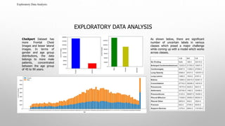
- 5. Exploratory Data Analysis: EXPLORATORY DATA ANALYSIS CheXpert Dataset has more Frontal Chest Images and lesser lateral images. In terms of gender and age group distributions, the data belongs to more male patients, concentrated between the age group of 45 to 90 years. As shown below, there are significant number of uncertain labels in various classes which posed a major challenge while coming up with a model which works across classes.
- 6. Exploratory Data Analysis: EXPLORATORY DATA ANALYSIS (CONT..) Below figure shows imbalance among the classes, along with significant number of uncertain labels in various classes. Further, We observed uneven distribution of data for positive, negative classes in 14 medical conditions during our analysis. Lots of missing or null values were observed along with uneven distribution in the 14 Medical Conditions.
- 7. ARCHITECTURAL FLOW DIAGRAM Training CXRs Chexpert Dataset consists of 224,316 check X-Ray images of 65,240 images for 14 Pathological observations. Test CXR Exploratory Data Analysis Exploratory Data Analysis will help to identify patterns, spot any anomalies, test given hypothesis, check assumptions with summary statistics and use of graphical representations. Data Preprocessing In this stage we are converting raw data into clean data set. Data preprocessing will include steps like data scaling and data augmentation. Feature Extraction Feature extraction by using Deep Learning Models (like LightNet-7, DenseNet121, Hybrid Model) to extract features from pre-processed image data. We have tried concept of Transfer Learning for this project. Train Classifier We are using Training data to fit the model where train & test ratio is 80:20. Predictive Model Model is now ready. It is capable to make predictions on unknown input x-ray image. Target Label Final output/pathology class that we are trying to predict. Pathology Label 14 Pathological observations. Performance Evaluation Evaluation of predictive model performance using various metrics
- 8. MODELLING APPROACH Following models were tried for the Project: 1. LightNet-7 : A 7 layered Deep Neural Network. 2. DenseNet121 from scratch. 3. DenseNet121 using pre-trained ImageNet weights. 4. Hybrid model with Random Forests (including extended features like Age-group, gender and type of image). 5. Hybrid model with AdaBoost (including extended features like Age-group, gender and type of image). 6. Hybrid model with XGBoost (including extended features like Age-group, gender and type of image).
- 9. Conv2D() -> Max Pool -> DropOut(0.5) Conv2D() -> Max Pool -> DropOut(0.5) Conv2D() -> Max Pool -> DropOut(0.5) Conv2D() -> Max Pool -> DropOut(0.5) Flatten() Dense(128,relu) -> DropOut(0.5) Dense(64,relu) -> DropOut(0.5) Dense(2,softmax) DEEP LEARNING MODELS USED – 7-LAYERED DNN (LIGHTNET-7*) conv2 D Max Pool Input Max Pool Max Pool Max Pool conv2 D conv2 D conv2 D Flattene d Fully Connected Neural Network + ReLU Activation With Dropout Prediction LightNet- 7
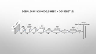
- 10. DEEP LEARNING MODELS USED – DENSENET121
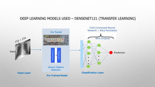
- 11. DEEP LEARNING MODELS USED – DENSENET121 (TRANSFER LEARNING) Pre Trained Generic Feature Detection Pre Trained Model Fully Connected Neural Network + ReLU Activation With Dropout Prediction Input Classification LayerInput Layer
- 12. HYBRID MODEL – DEEP NEURAL NETWORK WITH MACHINE LEARNING CLASSIFICATION ALGORITHM Pre Trained Generic Feature Detection Pre Trained Model Prediction Input Multiclass Classification Model Input Layer Machine Learning Models like AdaBoost, XGBoost or Random Forest
- 13. APPROACHES AND METHODOLOGY We will discuss below, our approach and methodology which we adopted for this project: We tried with Individual Class Wise Binary Classification, as training model together for all 14 Disease condition didn’t yield expected results. With this approach, we came across Imbalanced Class problem for almost all 14 Disease conditions. Further, we observed uneven distribution of data for positive, negative classes in 14 diseases along with missing or null values. To handle imbalanced class problem, we tried technique of up-sampling the minority class using SMOTE approach which improved our results significantly. We will now take a deep dive into other approaches used in the project : Data Pre-processing through Data Scaling and Data Augmentation. By Data Scaling, we created image tensors for Input images before feeding to DNN. Each Input Image Data was converted into Scaled Image Array with values between 0 and 1. An array list by consolidating all images was finally converted to numpy array. Also, to overcome issue of overfitting, Data Augmentation was done on input images to generate virtual images, by making transformations like flipping image, shearing and zooming. Feature Extraction using Transfer Learning, where we used weights of Pre-Trained DenseNet-121, trained on vast set of images. We used its initial layers to extract features from our images. We also tried a Hybrid Model, where DNN was used only for feature extraction and classical machine learning and ensemble method algorithms like Random Forest, AdaBoost and XGboost was used for Final classification.
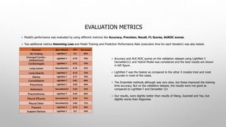
- 14. EVALUATION METRICS Model’s performance was evaluated by using different metrices like Accuracy, Precision, Recall, F1 Scores, AUROC scores. Two additional metrics Hamming Loss and Model Training and Prediction Performance Rate (execution time for each iteration) was also tested. Disease Best Model AUC Accuracy No Finding LightNet-7 0.5 84% Enlarged Cardio- mediastinum LightNet-7 0.79 79% Cardiomegaly LightNet-7 0.71 74% Lung Lesion DenseNet121 0.74 47% Lung Opacity LightNet-7 0.72 72% Edema LightNet-7 0.77 79% Consolidation LightNet-7 0.81 68% Pneumonia LightNet-7 0.69 64% Atelectasis DenseNet121 0.59 54% Pneumothorax LightNet-7 0.43 60% Pleural Effusion DenseNet121 0.69 72% Pleural Other DenseNet121 0.85 71% Fracture LightNet-7 0.73 45% Support Devices LightNet-7 0.5 54% Accuracy and AUC-ROC scores on the validation dataset using LightNet-7, DenseNet121 and Hybrid Model was considered and the best results are shown in left figure. LightNet-7 was the fastest as compared to the other 5 models tried and most accurate in most of the cases. The Ensemble methods although was very slow, but these improved the training time accuracy. But on the validation dataset, the results were not good as compared to LightNet-7 and DenseNet 121 Our results, were slightly better than results of Wang, Guendel and Yao, but slightly worse than Rajpurkar.
- 15. RESULTS AND CONCLUSION Through this project, we tried to come up with a framework to classify 14 thoracic diseases using CheXpert dataset, splitting it in 70-10-20 ratio. In this framework, we used pre-trained DenseNet121 for Feature Extraction. For Classification, we have experimented with CNN model, VGG16, Densenet121, ResNet models and the concept of Transfer Learning. We even tried classical Machine Learning approaches like Random Forests, AdaBoost and XGBoost for classification. For evaluating model's performance, we used metrics like Confusion matrices, Accuracy, Precision, Recall, F1 Scores, AUROC Scores and Hamming Loss although we presented just Accuracy and AUC in this presentation. Hybrid models are improving accuracy with more training but not improving AUC (especially on validation set) and they are slower than others As it was shown in slide before, our custom LightNet-7 worked better than others in most of the cases. DenseNet121 was the second best. LightNet-7 also proved to be faster to train as compared to other models.
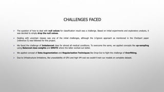
- 16. CHALLENGES FACED The question of how to deal with null values for classification result was a challenge. Based on initial experiments and exploratory analysis, it was decided to simply drop the null values. Dealing with uncertain classes was one of the initial challenges, although the U-Ignore approach as mentioned in the CheXpert paper (reference 5) was followed for this project. We faced the challenge of Imbalanced class for almost all medical conditions. To overcome the same, we applied concepts like up-sampling using Balanced class weights and SMOTE where the latter worked out better. We applied concept of Data Augmentation and Regularization Techniques like Drop-Out to fight the challenge of Overfitting. Due to Infrastructure limitations, like unavailability of GPU and high VM cost we couldn’t train our models on complete dataset.
- 17. IMPROVEMENTS AND FUTURE WORKS The SMOTE approach to handle imbalanced data was taking very long as it tries to oversample from the entire dataset. Instead we would try to come up with an algorithm which oversamples from random batches from the entire dataset. Another future improvement can be specific localization with the help of Regional CNN (R-CNN). We would like to try other approaches like U-One, U-Zero, U-SelfTrained mentioned in the CheXpert paper (reference 5) on the current models to handle uncertain labels and see which approach works better for the various disease classes.
- 18. THANK YOU! Questions? - Want to connect over LinkedIn ? - Or email me at: [email protected]
Editor's Notes
- #10: *LightNet-7- We named out 7-Layer DNN Model as LightNet-7