SlideShare a Scribd company logo
2
Most read
3
Most read
8
Most read
© 2023, IRJET | Impact Factor value: 8.226 | ISO 9001:2008 Certified Journal | Page 188
Brain Tumor Detection Using Deep Learning
Mr. Rijul Jain1, Mr. Karan Kumar Gupta2, Mr. Akshansh Kumar3, Ms. Kavita Saxena4
1,2,3 Department of Computer Science and Engineering Maharaja Agrasen Institute of Technology Delhi, India
4(Assistant Professor, CSE) Maharaja Agrasen Institute of Technology of Technology Delhi, India
------------------------------------------------------------------------------***------------------------------------------------------------------------
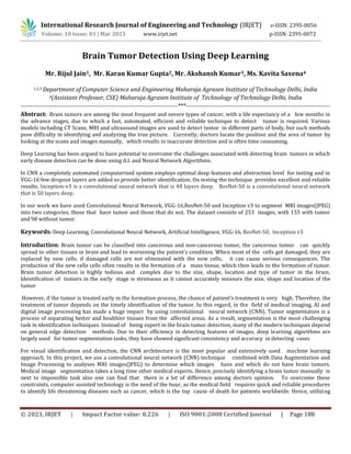
Abstract: Brain tumors are among the most frequent and severe types of cancer, with a life expectancy of a few months in
the advance stages, due to which a fast, automated, efficient and reliable technique to detect tumor is required. Various
models including CT Scans, MRI and ultrasound images are used to detect tumor in different parts of body, but such methods
pose difficulty in identifying and analyzing the true picture. Currently, doctors locate the position and the area of tumor by
looking at the scans and images manually, which results in inaccurate detection and is often time consuming.
Deep Learning has been argued to have potential to overcome the challenges associated with detecting brain tumors in which
early disease detection can be done using A.I. and Neural Network Algorithms.
In CNN a completely automated computerized system employs optimal deep features and abstraction level for testing and in
VGG-16 few dropout layers are added so provide better identification. On testing the technique provides excellent and reliable
results. Inception-v3 is a convolutional neural network that is 48 layers deep. ResNet-50 is a convolutional neural network
that is 50 layers deep.
In our work we have used Convolutional Neural Network, VGG-16,ResNet-50 and Inception v3 to segment MRI images(JPEG)
into two categories, those that have tumor and those that do not. The dataset consists of 253 images, with 155 with tumor
and 98 without tumor.
Keywords: Deep Learning, Convolutional Neural Network, Artificial Intelligence, VGG-16, ResNet-50, Inception v3
Introduction: Brain tumor can be classified into cancerous and non-cancerous tumor, the cancerous tumor can quickly
spread to other tissues in brain and lead to worsening the patient’s condition. When most of the cells get damaged, they are
replaced by new cells. if damaged cells are not eliminated with the new cells, it can cause serious consequences. The
production of the new cells cells often results in the formation of a mass tissue, which then leads to the formation of tumor.
Brain tumor detection is highly tedious and complex due to the size, shape, location and type of tumor in the brain.
Identification of tumors in the early stage is strenuous as it cannot accurately measure the size, shape and location of the
tumor
However, if the tumor is treated early in the formation process, the chance of patient’s treatment is very high. Therefore, the
treatment of tumor depends on the timely identification of the tumor. In this regard, in the field of medical imaging, AI and
digital image processing has made a huge impact by using convolutional neural network (CNN). Tumor segmentation is a
process of separating better and healthier tissues from the affected areas. As a result, segmentation is the most challenging
task in identification techniques. Instead of being expert in the brain tumor detection, many of the modern techniques depend
on general edge detection methods. Due to their efficiency in detecting features of images, deep learning algorithms are
largely used for tumor segmentation tasks, they have showed significant consistency and accuracy in detecting cases
For visual identification and detection, the CNN architecture is the most popular and extensively used machine learning
approach. In this project, we use a convolutional neural network (CNN) technique combined with Data Augmentation and
Image Processing to analyses MRI images(JPEG) to determine which images have and which do not have brain tumors.
Medical image segmentation takes a long time other medical experts. Hence, precisely identifying a brain tumor manually is
next to impossible task also one can find that there is a lot of difference among doctors opinion. To overcome these
constraints, computer assisted technology is the need of the hour, as the medical field requires quick and reliable procedures
to identify life threatening diseases such as cancer, which is the top cause of death for patients worldwide. Hence, utilizing
International Research Journal of Engineering and Technology (IRJET) e-ISSN: 2395-0056
Volume: 10 Issue: 03 | Mar 2023 www.irjet.net p-ISSN: 2395-0072
International Research Journal of Engineering and Technology (IRJET) e-ISSN: 2395-0056
Volume: 10 Issue: 03 | Mar 2023 www.irjet.net p-ISSN: 2395-0072
© 2023, IRJET | Impact Factor value: 8.226 | ISO 9001:2008 Certified Journal | Page 189
Brain MRI Images(JPEG), we present a method for classifying MRI images(JPEG) into those with and without the presence of a
brain tumors utilizing a data augmentation strategy and a convolutional neural network model in our study.
Proposed System: Edge detection is a process of finding and examining certain disruptions in an image. The disruptions are
sudden changes in the information stored in image which identifies boundaries of objects in a scene. Currently, there are
many edge detection techniques available which are used to identify specific types of edges. The aim is to reduce the
information stored in the image while conserving the required properties
Efficient edge detection mechanism is used in the project
Fig.1 Edge detection
We propose a method that is sequential in its operation. It uses techniques such as convolutional neural network, max
pooling, flattening and a dropout layer to avoid over-fitting. We use transfer learning to assign weights to our features. The
MRI image(JPEG) sequentially goes through the above stated layers and feature extraction is performed.DICOM images is
large in size when we use to it to train model parameter increase so to decrease parameter we use dicom converted jpeg
images from Kaggle. The MRI images(JPEG) have been divided into training and testing parts where the first part is used for
further training of a VGG-16 CNN, ResNet-50,Inception v3.
Algorithm demonstrates the step-by-step working process of the proposed system.
International Research Journal of Engineering and Technology (IRJET) e-ISSN: 2395-0056
Volume: 10 Issue: 03 | Mar 2023 www.irjet.net p-ISSN: 2395-0072
© 2023, IRJET | Impact Factor value: 8.226 | ISO 9001:2008 Certified Journal | Page 190
Fig. 2 Flowchart
Literature Review: The lump in the brain tumor can be present at different locations and shapes, during the time of
segmentation . Brain tumors are segmented using the MRF system. Generative models successfully generalize hidden data
with some constraints in the training step. Without the use of a specific model, these strategies can be employed to
comprehend the pattern of a brain tumor. The distribution of identical and independent voxels on the ground of context
factors is constantly taken into account in these styles. As a result, some small or isolated clusters of voxels may be
distributed incorrectly into the wrong class, generally in anatomically and physiologically incongruent places. To avoid
these issues, numerous experimenters used probabilistic predictions to fit neighborhood information into a Conditional
Random Field (CRF) classifier. Deep CNN models are used to automatically learn scales of complex data attributes.
In this work we have enforced a pre-trained and advanced interpretation of VGG-16 Convolutional Neural Network (CNN) to
categorize brain tumor images taken with camera into two types that is cancerous and normal images. Although the network
model does not need feature extraction to apply a small quantum, training CNN architecture is tough and tedious since it
needs a dataset for testing and training before the structure is ready for classification, which is not always available. In
addition, hardware is required for computing the massive factor for large images. . Since DL Algorithms can effectively
express critical relations without demanding a wide variety of equipment, they are a remarkable development in ML . As a
result, they evolved fleetly to serve as a blessing in several health informatics fields, such as informatics, healthcare analytics,
and pattern identification.
Methodology Result and Discussion: The dataset used in the design is Brain MRI Images(JPEG) for Brain Tumor
Identification , which is easily available on Kaggle. The dataset includes 253 images of brain MRI ,out of which 155 have
tumour and 98 images do not have tumour. Our dataset was divided into three parts for training, validation, and testing. The
International Research Journal of Engineering and Technology (IRJET) e-ISSN: 2395-0056
Volume: 10 Issue: 03 | Mar 2023 www.irjet.net p-ISSN: 2395-0072
© 2023, IRJET | Impact Factor value: 8.226 | ISO 9001:2008 Certified Journal | Page 191
training data is used to learn the model and get used to the features, while the validation data is used to estimate the model
and interpret its parameters.
The validation is implemented using Cross-Validation. Our model’s final evaluation will be on the ground of test data.
In some cases, the areas of fat in the images are incorrectly identified as tumor, or the tumors may not be seen by the doctor;
the most exact treatment is completely depended on the doctor’s skill. In this paper, the CNN has been used for Brain tumor
detection using some images. There were some margins of the images gathered from the imaging centers, which were
removed to provide a high resolution view of the image One of the main reasons for using the feature extraction technique
was to increase the accuracy of the network.
Fig. 3 Data Set
International Research Journal of Engineering and Technology (IRJET) e-ISSN: 2395-0056
Volume: 10 Issue: 03 | Mar 2023 www.irjet.net p-ISSN: 2395-0072
© 2023, IRJET | Impact Factor value: 8.226 | ISO 9001:2008 Certified Journal | Page 192
International Research Journal of Engineering and Technology (IRJET) e-ISSN: 2395-0056
Volume: 10 Issue: 03 | Mar 2023 www.irjet.net p-ISSN: 2395-0072
© 2023, IRJET | Impact Factor value: 8.226 | ISO 9001:2008 Certified Journal | Page 193
International Research Journal of Engineering and Technology (IRJET) e-ISSN: 2395-0056
Volume: 10 Issue: 03 | Mar 2023 www.irjet.net p-ISSN: 2395-0072
© 2023, IRJET | Impact Factor value: 8.226 | ISO 9001:2008 Certified Journal | Page 194
Images were reshaped into 244×244 size and passed through a convolutional layer with three filters. Subsequently, the output
passes through maxpool and flattening layers. The model has been trained with 30,120 epochs.
The essential variables stated for the given model showed that it was effective in finding original tumor areas, while avoiding
false ones. Most current techniques focus on the entire tumor region, resulting in incorrect measurements for core and
augment regions. The methods proposed in the document should include statistical and deep learningbased technology, with
CNN dealing with task complexity. Due to the clinical significance of the tumor detection problem, time constraints, sensitivity,
and accuracy are essential. The results validated the efficiency and efficacy of the proposed models, especially in terms of
fundamental and augmenting regions of some values, where it outperformed previous methods by a wide margin.
MODEL ACCURACY PRECISION RECALL F1 SCORE
CNN 50% 50% 100% 66.7%
Inception v3 60% 57.1% 80% 66.7%
ResNet-50 90% 100% 80% 88.9%
VGG-16 90% 100% 80% 88.9%
Fig. 4 Result Table
International Research Journal of Engineering and Technology (IRJET) e-ISSN: 2395-0056
Volume: 10 Issue: 03 | Mar 2023 www.irjet.net p-ISSN: 2395-0072
© 2023, IRJET | Impact Factor value: 8.226 | ISO 9001:2008 Certified Journal | Page 195
Conclusion and Future Scope: In this project, we used MRI images(JPEG) of the brain to divide into two types, one having
tumor and the other not having tumor. We executed a sequential model where the images were reshaped to 244 x 244 then
the convolutional layer, max-pooling layer, flattening is performed on those images to convert it to a vector form. Hence, we
used ImageNet dataset which contains a large number of medical pictures, which helps in feature enhancement. The
architecture used is VGG-16, ResNet-50, Inception v3 which is further improved by using a dropout layer. When compared to
manual detection done by doctors or clinical physicians, the results of the experiments on different pictures show that the
analysis for brain tumour detection is quick and efficient. Our research show that the described way can help in the accurate
and timely detection of brain tumours, as well as it can locate the exact region of tumor. As a result, the given method is
essential for identifying tumours from images. According to our research, the given method is essential for taking the correct
decision by medical experts
The given method can be expanded for better classification in future research. These methods can efficiently treat other types
of tumors and disorders. This model can be used during operations for searching and locating the exact region of tumor.
Detecting tumours in the operation theatre could be done in real time situations. Future research can make use of methods to
acquire the nearest location of infected area in the brain to isolate them from unaffected parts, testing the efficiency of neural
network, improved models could be done in the near future.
References:
[1] Nilesh Bhaskarrao Bahadure, Arun Kumar Ray, and Har Pal Thethi, “Image Analysis for MRI Based Brain Tumor Detection
and Feature Extraction Using Biologically Inspired BWT and SVM”, 2017
[2] Luxit Kapoor, Sanjeev Thakur “A Survey on Brain Tumor Detection Using Image Processing Techniques”, 2017
[3] Praveen Gamage “Identification of Brain Tumor using Image Processing Techniques”,2017
[4] Deepa, Akansha Singh “Review of Brain Tumor Detection from MRI Images”, 2016
[5] Devendra Somwanshi , Ashutosh Kumar, Pratima Sharma, Deepika Joshi “An efficient Brain Tumor Detection from MRI
Images using Entropy Measures ”, 2016
[6] A. Demirhan, M. Toru, and I. Guler, “Segmentation of tumor and edema along with healthy tissues of brain using wavelets
and neural networks”, 2015
[7] Yaqub, M.; Feng, J.; Zia, M.S.; Arshid, K.; Jia, K.; Rehman, Z.U.; Mehmood, A. “State-of-the-art CNN optimizer for brain tumor
segmentation in magnetic resonance images”, 2020
[8] Amin, J.; Sharif, M.; Yasmin, M.; Fernandes, S.L. “A distinctive approach in brain tumor detection and classification using
M.R.I. Pattern Recognit. Lett.”, 2020

More Related Content

Similar to Brain Tumor Detection Using Deep Learning (20)

PDF
Brain tumor classification in magnetic resonance imaging images using convol...
IJECEIAES
 
PDF
IRJET- An Efficient Brain Tumor Detection System using Automatic Segmenta...
IRJET Journal
 
PPTX
Your wish you too happy birthday to you too
halderdhrubo6
 
PDF
AI Based Approach for Classification of MultiGrade Tumour in Human Brain
IRJET Journal
 
PDF
IRJET- Brain Tumor Detection using Convolutional Neural Network
IRJET Journal
 
PDF
DIRECTIONAL CLASSIFICATION OF BRAIN TUMOR IMAGES FROM MRI USING CNN-BASED DEE...
IRJET Journal
 
PDF
A Comparative Study of Various Machine Learning Techniques for Brain Tumor De...
IRJET Journal
 
PDF
Brain Tumor Detection using Neural Network
ijtsrd
 
PDF
A Review Paper on Automated Brain Tumor Detection
IRJET Journal
 
PDF
BRAIN TUMOR DETECTION USING CNN & ML TECHNIQUES
IRJET Journal
 
PPTX
Major Project Internship Review-2 PPT Template-WS 23-24.pptx
HIMANSHUKUMARCse2020
 
PDF
DEEP LEARNING METHODS FOR IDENTIFYING BRAIN TUMORS.pdf
sspub2000
 
PDF
Brain tumor detection using Region-based Convolutional Neural Network and PSO
IRJET Journal
 
PDF
IRJET- Image Classification using Deep Learning Neural Networks for Brain...
IRJET Journal
 
PDF
A review on detecting brain tumors using deep learning and magnetic resonanc...
IJECEIAES
 
PPTX
Brain Tumor Detection Using Deep Neural Network.pptx
AbdulRehman417114
 
PDF
Brain tumor classification using artificial neural network on mri images
eSAT Journals
 
PPTX
Brain Tumor Detection Using Deep Learning ppt new made.pptx
vikyt2211
 
PDF
NEUROSENTINEL PRODIGY (Brain Tumor) using CNN
vivatechijri
 
PPT
batch 14.ppt aitam useful ppt for students
Himabindu905359
 
Brain tumor classification in magnetic resonance imaging images using convol...
IJECEIAES
 
IRJET- An Efficient Brain Tumor Detection System using Automatic Segmenta...
IRJET Journal
 
Your wish you too happy birthday to you too
halderdhrubo6
 
AI Based Approach for Classification of MultiGrade Tumour in Human Brain
IRJET Journal
 
IRJET- Brain Tumor Detection using Convolutional Neural Network
IRJET Journal
 
DIRECTIONAL CLASSIFICATION OF BRAIN TUMOR IMAGES FROM MRI USING CNN-BASED DEE...
IRJET Journal
 
A Comparative Study of Various Machine Learning Techniques for Brain Tumor De...
IRJET Journal
 
Brain Tumor Detection using Neural Network
ijtsrd
 
A Review Paper on Automated Brain Tumor Detection
IRJET Journal
 
BRAIN TUMOR DETECTION USING CNN & ML TECHNIQUES
IRJET Journal
 
Major Project Internship Review-2 PPT Template-WS 23-24.pptx
HIMANSHUKUMARCse2020
 
DEEP LEARNING METHODS FOR IDENTIFYING BRAIN TUMORS.pdf
sspub2000
 
Brain tumor detection using Region-based Convolutional Neural Network and PSO
IRJET Journal
 
IRJET- Image Classification using Deep Learning Neural Networks for Brain...
IRJET Journal
 
A review on detecting brain tumors using deep learning and magnetic resonanc...
IJECEIAES
 
Brain Tumor Detection Using Deep Neural Network.pptx
AbdulRehman417114
 
Brain tumor classification using artificial neural network on mri images
eSAT Journals
 
Brain Tumor Detection Using Deep Learning ppt new made.pptx
vikyt2211
 
NEUROSENTINEL PRODIGY (Brain Tumor) using CNN
vivatechijri
 
batch 14.ppt aitam useful ppt for students
Himabindu905359
 

More from IRJET Journal (20)

PDF
Enhanced heart disease prediction using SKNDGR ensemble Machine Learning Model
IRJET Journal
 
PDF
Utilizing Biomedical Waste for Sustainable Brick Manufacturing: A Novel Appro...
IRJET Journal
 
PDF
Kiona – A Smart Society Automation Project
IRJET Journal
 
PDF
DESIGN AND DEVELOPMENT OF BATTERY THERMAL MANAGEMENT SYSTEM USING PHASE CHANG...
IRJET Journal
 
PDF
Invest in Innovation: Empowering Ideas through Blockchain Based Crowdfunding
IRJET Journal
 
PDF
SPACE WATCH YOUR REAL-TIME SPACE INFORMATION HUB
IRJET Journal
 
PDF
A Review on Influence of Fluid Viscous Damper on The Behaviour of Multi-store...
IRJET Journal
 
PDF
Wireless Arduino Control via Mobile: Eliminating the Need for a Dedicated Wir...
IRJET Journal
 
PDF
Explainable AI(XAI) using LIME and Disease Detection in Mango Leaf by Transfe...
IRJET Journal
 
PDF
BRAIN TUMOUR DETECTION AND CLASSIFICATION
IRJET Journal
 
PDF
The Project Manager as an ambassador of the contract. The case of NEC4 ECC co...
IRJET Journal
 
PDF
"Enhanced Heat Transfer Performance in Shell and Tube Heat Exchangers: A CFD ...
IRJET Journal
 
PDF
Advancements in CFD Analysis of Shell and Tube Heat Exchangers with Nanofluid...
IRJET Journal
 
PDF
Breast Cancer Detection using Computer Vision
IRJET Journal
 
PDF
Auto-Charging E-Vehicle with its battery Management.
IRJET Journal
 
PDF
Analysis of high energy charge particle in the Heliosphere
IRJET Journal
 
PDF
A Novel System for Recommending Agricultural Crops Using Machine Learning App...
IRJET Journal
 
PDF
Auto-Charging E-Vehicle with its battery Management.
IRJET Journal
 
PDF
Analysis of high energy charge particle in the Heliosphere
IRJET Journal
 
PDF
Wireless Arduino Control via Mobile: Eliminating the Need for a Dedicated Wir...
IRJET Journal
 
Enhanced heart disease prediction using SKNDGR ensemble Machine Learning Model
IRJET Journal
 
Utilizing Biomedical Waste for Sustainable Brick Manufacturing: A Novel Appro...
IRJET Journal
 
Kiona – A Smart Society Automation Project
IRJET Journal
 
DESIGN AND DEVELOPMENT OF BATTERY THERMAL MANAGEMENT SYSTEM USING PHASE CHANG...
IRJET Journal
 
Invest in Innovation: Empowering Ideas through Blockchain Based Crowdfunding
IRJET Journal
 
SPACE WATCH YOUR REAL-TIME SPACE INFORMATION HUB
IRJET Journal
 
A Review on Influence of Fluid Viscous Damper on The Behaviour of Multi-store...
IRJET Journal
 
Wireless Arduino Control via Mobile: Eliminating the Need for a Dedicated Wir...
IRJET Journal
 
Explainable AI(XAI) using LIME and Disease Detection in Mango Leaf by Transfe...
IRJET Journal
 
BRAIN TUMOUR DETECTION AND CLASSIFICATION
IRJET Journal
 
The Project Manager as an ambassador of the contract. The case of NEC4 ECC co...
IRJET Journal
 
"Enhanced Heat Transfer Performance in Shell and Tube Heat Exchangers: A CFD ...
IRJET Journal
 
Advancements in CFD Analysis of Shell and Tube Heat Exchangers with Nanofluid...
IRJET Journal
 
Breast Cancer Detection using Computer Vision
IRJET Journal
 
Auto-Charging E-Vehicle with its battery Management.
IRJET Journal
 
Analysis of high energy charge particle in the Heliosphere
IRJET Journal
 
A Novel System for Recommending Agricultural Crops Using Machine Learning App...
IRJET Journal
 
Auto-Charging E-Vehicle with its battery Management.
IRJET Journal
 
Analysis of high energy charge particle in the Heliosphere
IRJET Journal
 
Wireless Arduino Control via Mobile: Eliminating the Need for a Dedicated Wir...
IRJET Journal
 
Ad

Recently uploaded (20)

PPTX
waterconservation-211128055737.pptx Jaswanth
SandulaAnilBabu
 
PDF
Detailed manufacturing Engineering and technology notes
VIKKYsing
 
PDF
Ethics and Trustworthy AI in Healthcare – Governing Sensitive Data, Profiling...
AlqualsaDIResearchGr
 
PDF
BioSensors glucose monitoring, cholestrol
nabeehasahar1
 
PDF
Passive building design opening approach
Dr-Fatima Um Mgdad
 
PPTX
Electron Beam Machining for Production Process
Rajshahi University of Engineering & Technology(RUET), Bangladesh
 
PDF
Set Relation Function Practice session 24.05.2025.pdf
DrStephenStrange4
 
PDF
1_ISO Certifications by Indian Industrial Standards Organisation.pdf
muhammad2010960
 
PDF
Statistical Data Analysis Using SPSS Software
shrikrishna kesharwani
 
PPTX
artificial intelligence applications in Geomatics
NawrasShatnawi1
 
PDF
Non Text Magic Studio Magic Design for Presentations L&P.pdf
rajpal7872
 
PPTX
Types of Bearing_Specifications_PPT.pptx
PranjulAgrahariAkash
 
PDF
POWER PLANT ENGINEERING (R17A0326).pdf..
haneefachosa123
 
PDF
Natural Language processing and web deigning notes
AnithaSakthivel3
 
PPTX
File Strucutres and Access in Data Structures
mwaslam2303
 
PPTX
Pharmaceuticals and fine chemicals.pptxx
jaypa242004
 
PPTX
MPMC_Module-2 xxxxxxxxxxxxxxxxxxxxx.pptx
ShivanshVaidya5
 
PPTX
Coding about python and MySQL connectivity
inderjitsingh1985as
 
PPT
Tiles.ppt The purpose of a floor is to provide a level surface capable of sup...
manojaioe
 
PPT
Oxygen Co2 Transport in the Lungs(Exchange og gases)
SUNDERLINSHIBUD
 
waterconservation-211128055737.pptx Jaswanth
SandulaAnilBabu
 
Detailed manufacturing Engineering and technology notes
VIKKYsing
 
Ethics and Trustworthy AI in Healthcare – Governing Sensitive Data, Profiling...
AlqualsaDIResearchGr
 
BioSensors glucose monitoring, cholestrol
nabeehasahar1
 
Passive building design opening approach
Dr-Fatima Um Mgdad
 
Electron Beam Machining for Production Process
Rajshahi University of Engineering & Technology(RUET), Bangladesh
 
Set Relation Function Practice session 24.05.2025.pdf
DrStephenStrange4
 
1_ISO Certifications by Indian Industrial Standards Organisation.pdf
muhammad2010960
 
Statistical Data Analysis Using SPSS Software
shrikrishna kesharwani
 
artificial intelligence applications in Geomatics
NawrasShatnawi1
 
Non Text Magic Studio Magic Design for Presentations L&P.pdf
rajpal7872
 
Types of Bearing_Specifications_PPT.pptx
PranjulAgrahariAkash
 
POWER PLANT ENGINEERING (R17A0326).pdf..
haneefachosa123
 
Natural Language processing and web deigning notes
AnithaSakthivel3
 
File Strucutres and Access in Data Structures
mwaslam2303
 
Pharmaceuticals and fine chemicals.pptxx
jaypa242004
 
MPMC_Module-2 xxxxxxxxxxxxxxxxxxxxx.pptx
ShivanshVaidya5
 
Coding about python and MySQL connectivity
inderjitsingh1985as
 
Tiles.ppt The purpose of a floor is to provide a level surface capable of sup...
manojaioe
 
Oxygen Co2 Transport in the Lungs(Exchange og gases)
SUNDERLINSHIBUD
 
Ad

Brain Tumor Detection Using Deep Learning

  • 1. © 2023, IRJET | Impact Factor value: 8.226 | ISO 9001:2008 Certified Journal | Page 188 Brain Tumor Detection Using Deep Learning Mr. Rijul Jain1, Mr. Karan Kumar Gupta2, Mr. Akshansh Kumar3, Ms. Kavita Saxena4 1,2,3 Department of Computer Science and Engineering Maharaja Agrasen Institute of Technology Delhi, India 4(Assistant Professor, CSE) Maharaja Agrasen Institute of Technology of Technology Delhi, India ------------------------------------------------------------------------------***------------------------------------------------------------------------ Abstract: Brain tumors are among the most frequent and severe types of cancer, with a life expectancy of a few months in the advance stages, due to which a fast, automated, efficient and reliable technique to detect tumor is required. Various models including CT Scans, MRI and ultrasound images are used to detect tumor in different parts of body, but such methods pose difficulty in identifying and analyzing the true picture. Currently, doctors locate the position and the area of tumor by looking at the scans and images manually, which results in inaccurate detection and is often time consuming. Deep Learning has been argued to have potential to overcome the challenges associated with detecting brain tumors in which early disease detection can be done using A.I. and Neural Network Algorithms. In CNN a completely automated computerized system employs optimal deep features and abstraction level for testing and in VGG-16 few dropout layers are added so provide better identification. On testing the technique provides excellent and reliable results. Inception-v3 is a convolutional neural network that is 48 layers deep. ResNet-50 is a convolutional neural network that is 50 layers deep. In our work we have used Convolutional Neural Network, VGG-16,ResNet-50 and Inception v3 to segment MRI images(JPEG) into two categories, those that have tumor and those that do not. The dataset consists of 253 images, with 155 with tumor and 98 without tumor. Keywords: Deep Learning, Convolutional Neural Network, Artificial Intelligence, VGG-16, ResNet-50, Inception v3 Introduction: Brain tumor can be classified into cancerous and non-cancerous tumor, the cancerous tumor can quickly spread to other tissues in brain and lead to worsening the patient’s condition. When most of the cells get damaged, they are replaced by new cells. if damaged cells are not eliminated with the new cells, it can cause serious consequences. The production of the new cells cells often results in the formation of a mass tissue, which then leads to the formation of tumor. Brain tumor detection is highly tedious and complex due to the size, shape, location and type of tumor in the brain. Identification of tumors in the early stage is strenuous as it cannot accurately measure the size, shape and location of the tumor However, if the tumor is treated early in the formation process, the chance of patient’s treatment is very high. Therefore, the treatment of tumor depends on the timely identification of the tumor. In this regard, in the field of medical imaging, AI and digital image processing has made a huge impact by using convolutional neural network (CNN). Tumor segmentation is a process of separating better and healthier tissues from the affected areas. As a result, segmentation is the most challenging task in identification techniques. Instead of being expert in the brain tumor detection, many of the modern techniques depend on general edge detection methods. Due to their efficiency in detecting features of images, deep learning algorithms are largely used for tumor segmentation tasks, they have showed significant consistency and accuracy in detecting cases For visual identification and detection, the CNN architecture is the most popular and extensively used machine learning approach. In this project, we use a convolutional neural network (CNN) technique combined with Data Augmentation and Image Processing to analyses MRI images(JPEG) to determine which images have and which do not have brain tumors. Medical image segmentation takes a long time other medical experts. Hence, precisely identifying a brain tumor manually is next to impossible task also one can find that there is a lot of difference among doctors opinion. To overcome these constraints, computer assisted technology is the need of the hour, as the medical field requires quick and reliable procedures to identify life threatening diseases such as cancer, which is the top cause of death for patients worldwide. Hence, utilizing International Research Journal of Engineering and Technology (IRJET) e-ISSN: 2395-0056 Volume: 10 Issue: 03 | Mar 2023 www.irjet.net p-ISSN: 2395-0072
  • 2. International Research Journal of Engineering and Technology (IRJET) e-ISSN: 2395-0056 Volume: 10 Issue: 03 | Mar 2023 www.irjet.net p-ISSN: 2395-0072 © 2023, IRJET | Impact Factor value: 8.226 | ISO 9001:2008 Certified Journal | Page 189 Brain MRI Images(JPEG), we present a method for classifying MRI images(JPEG) into those with and without the presence of a brain tumors utilizing a data augmentation strategy and a convolutional neural network model in our study. Proposed System: Edge detection is a process of finding and examining certain disruptions in an image. The disruptions are sudden changes in the information stored in image which identifies boundaries of objects in a scene. Currently, there are many edge detection techniques available which are used to identify specific types of edges. The aim is to reduce the information stored in the image while conserving the required properties Efficient edge detection mechanism is used in the project Fig.1 Edge detection We propose a method that is sequential in its operation. It uses techniques such as convolutional neural network, max pooling, flattening and a dropout layer to avoid over-fitting. We use transfer learning to assign weights to our features. The MRI image(JPEG) sequentially goes through the above stated layers and feature extraction is performed.DICOM images is large in size when we use to it to train model parameter increase so to decrease parameter we use dicom converted jpeg images from Kaggle. The MRI images(JPEG) have been divided into training and testing parts where the first part is used for further training of a VGG-16 CNN, ResNet-50,Inception v3. Algorithm demonstrates the step-by-step working process of the proposed system.
  • 3. International Research Journal of Engineering and Technology (IRJET) e-ISSN: 2395-0056 Volume: 10 Issue: 03 | Mar 2023 www.irjet.net p-ISSN: 2395-0072 © 2023, IRJET | Impact Factor value: 8.226 | ISO 9001:2008 Certified Journal | Page 190 Fig. 2 Flowchart Literature Review: The lump in the brain tumor can be present at different locations and shapes, during the time of segmentation . Brain tumors are segmented using the MRF system. Generative models successfully generalize hidden data with some constraints in the training step. Without the use of a specific model, these strategies can be employed to comprehend the pattern of a brain tumor. The distribution of identical and independent voxels on the ground of context factors is constantly taken into account in these styles. As a result, some small or isolated clusters of voxels may be distributed incorrectly into the wrong class, generally in anatomically and physiologically incongruent places. To avoid these issues, numerous experimenters used probabilistic predictions to fit neighborhood information into a Conditional Random Field (CRF) classifier. Deep CNN models are used to automatically learn scales of complex data attributes. In this work we have enforced a pre-trained and advanced interpretation of VGG-16 Convolutional Neural Network (CNN) to categorize brain tumor images taken with camera into two types that is cancerous and normal images. Although the network model does not need feature extraction to apply a small quantum, training CNN architecture is tough and tedious since it needs a dataset for testing and training before the structure is ready for classification, which is not always available. In addition, hardware is required for computing the massive factor for large images. . Since DL Algorithms can effectively express critical relations without demanding a wide variety of equipment, they are a remarkable development in ML . As a result, they evolved fleetly to serve as a blessing in several health informatics fields, such as informatics, healthcare analytics, and pattern identification. Methodology Result and Discussion: The dataset used in the design is Brain MRI Images(JPEG) for Brain Tumor Identification , which is easily available on Kaggle. The dataset includes 253 images of brain MRI ,out of which 155 have tumour and 98 images do not have tumour. Our dataset was divided into three parts for training, validation, and testing. The
  • 4. International Research Journal of Engineering and Technology (IRJET) e-ISSN: 2395-0056 Volume: 10 Issue: 03 | Mar 2023 www.irjet.net p-ISSN: 2395-0072 © 2023, IRJET | Impact Factor value: 8.226 | ISO 9001:2008 Certified Journal | Page 191 training data is used to learn the model and get used to the features, while the validation data is used to estimate the model and interpret its parameters. The validation is implemented using Cross-Validation. Our model’s final evaluation will be on the ground of test data. In some cases, the areas of fat in the images are incorrectly identified as tumor, or the tumors may not be seen by the doctor; the most exact treatment is completely depended on the doctor’s skill. In this paper, the CNN has been used for Brain tumor detection using some images. There were some margins of the images gathered from the imaging centers, which were removed to provide a high resolution view of the image One of the main reasons for using the feature extraction technique was to increase the accuracy of the network. Fig. 3 Data Set
  • 5. International Research Journal of Engineering and Technology (IRJET) e-ISSN: 2395-0056 Volume: 10 Issue: 03 | Mar 2023 www.irjet.net p-ISSN: 2395-0072 © 2023, IRJET | Impact Factor value: 8.226 | ISO 9001:2008 Certified Journal | Page 192
  • 6. International Research Journal of Engineering and Technology (IRJET) e-ISSN: 2395-0056 Volume: 10 Issue: 03 | Mar 2023 www.irjet.net p-ISSN: 2395-0072 © 2023, IRJET | Impact Factor value: 8.226 | ISO 9001:2008 Certified Journal | Page 193
  • 7. International Research Journal of Engineering and Technology (IRJET) e-ISSN: 2395-0056 Volume: 10 Issue: 03 | Mar 2023 www.irjet.net p-ISSN: 2395-0072 © 2023, IRJET | Impact Factor value: 8.226 | ISO 9001:2008 Certified Journal | Page 194 Images were reshaped into 244×244 size and passed through a convolutional layer with three filters. Subsequently, the output passes through maxpool and flattening layers. The model has been trained with 30,120 epochs. The essential variables stated for the given model showed that it was effective in finding original tumor areas, while avoiding false ones. Most current techniques focus on the entire tumor region, resulting in incorrect measurements for core and augment regions. The methods proposed in the document should include statistical and deep learningbased technology, with CNN dealing with task complexity. Due to the clinical significance of the tumor detection problem, time constraints, sensitivity, and accuracy are essential. The results validated the efficiency and efficacy of the proposed models, especially in terms of fundamental and augmenting regions of some values, where it outperformed previous methods by a wide margin. MODEL ACCURACY PRECISION RECALL F1 SCORE CNN 50% 50% 100% 66.7% Inception v3 60% 57.1% 80% 66.7% ResNet-50 90% 100% 80% 88.9% VGG-16 90% 100% 80% 88.9% Fig. 4 Result Table
  • 8. International Research Journal of Engineering and Technology (IRJET) e-ISSN: 2395-0056 Volume: 10 Issue: 03 | Mar 2023 www.irjet.net p-ISSN: 2395-0072 © 2023, IRJET | Impact Factor value: 8.226 | ISO 9001:2008 Certified Journal | Page 195 Conclusion and Future Scope: In this project, we used MRI images(JPEG) of the brain to divide into two types, one having tumor and the other not having tumor. We executed a sequential model where the images were reshaped to 244 x 244 then the convolutional layer, max-pooling layer, flattening is performed on those images to convert it to a vector form. Hence, we used ImageNet dataset which contains a large number of medical pictures, which helps in feature enhancement. The architecture used is VGG-16, ResNet-50, Inception v3 which is further improved by using a dropout layer. When compared to manual detection done by doctors or clinical physicians, the results of the experiments on different pictures show that the analysis for brain tumour detection is quick and efficient. Our research show that the described way can help in the accurate and timely detection of brain tumours, as well as it can locate the exact region of tumor. As a result, the given method is essential for identifying tumours from images. According to our research, the given method is essential for taking the correct decision by medical experts The given method can be expanded for better classification in future research. These methods can efficiently treat other types of tumors and disorders. This model can be used during operations for searching and locating the exact region of tumor. Detecting tumours in the operation theatre could be done in real time situations. Future research can make use of methods to acquire the nearest location of infected area in the brain to isolate them from unaffected parts, testing the efficiency of neural network, improved models could be done in the near future. References: [1] Nilesh Bhaskarrao Bahadure, Arun Kumar Ray, and Har Pal Thethi, “Image Analysis for MRI Based Brain Tumor Detection and Feature Extraction Using Biologically Inspired BWT and SVM”, 2017 [2] Luxit Kapoor, Sanjeev Thakur “A Survey on Brain Tumor Detection Using Image Processing Techniques”, 2017 [3] Praveen Gamage “Identification of Brain Tumor using Image Processing Techniques”,2017 [4] Deepa, Akansha Singh “Review of Brain Tumor Detection from MRI Images”, 2016 [5] Devendra Somwanshi , Ashutosh Kumar, Pratima Sharma, Deepika Joshi “An efficient Brain Tumor Detection from MRI Images using Entropy Measures ”, 2016 [6] A. Demirhan, M. Toru, and I. Guler, “Segmentation of tumor and edema along with healthy tissues of brain using wavelets and neural networks”, 2015 [7] Yaqub, M.; Feng, J.; Zia, M.S.; Arshid, K.; Jia, K.; Rehman, Z.U.; Mehmood, A. “State-of-the-art CNN optimizer for brain tumor segmentation in magnetic resonance images”, 2020 [8] Amin, J.; Sharif, M.; Yasmin, M.; Fernandes, S.L. “A distinctive approach in brain tumor detection and classification using M.R.I. Pattern Recognit. Lett.”, 2020