Component Preparation methods & techniques.
Download as PPTX, PDF0 likes691 views

The document discusses the modern methods of blood component preparation, emphasizing the separation of blood into specific components such as red cells, plasma, and platelets using centrifugation techniques based on specific gravity. Various centrifuge types, anticoagulants, and additive solutions used in preserving blood components are detailed, along with storage protocols for red blood cells and platelets. Additionally, it covers the preparation and storage of cryoprecipitate and fresh frozen plasma, outlining their respective shelf lives and transport conditions.


Component Preparation methods & techniques.
- 1. Component Preparation Dr. P K Maharana
- 2. Blood & its components.
- 3. Component Preparation Present concept is to transfuse the specific components of the blood instead of transfusing whole blood contrast to earlier practice. A single unit of blood collected can be segregated to different components like red cells, plasma, platelets & cryoprecipitate, which can be of use for four beneficiaries.
- 4. Preparation of Blood components • Blood is a mixture of cells and plasma. • Basing on the specific gravity of different components they can be separated in to individual components. Centrifuge is the device to spin and segregate different components according to their specific gravity.
- 5. Specific gravity of different cellular components Due to different specific gravity of cellular components, they can be separated by centrifuging at different centrifugal force (in g) for different time. Specific Gravity of different components listed below. Name of Component Specific Gravity • Whole blood 1.053 • Red Cells 1.08 – 1.09 • Platelet 1.03 – 1. 04 • Plasma 1.02 – 1. 03
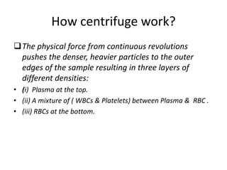
- 6. How centrifuge work? The physical force from continuous revolutions pushes the denser, heavier particles to the outer edges of the sample resulting in three layers of different densities: • (i) Plasma at the top. • (ii) A mixture of ( WBCs & Platelets) between Plasma & RBC . • (iii) RBCs at the bottom.
- 7. The 3 Layers On Sedimentation or Centrifugation blood separates to three different layers. • 1.Plasma: Occupies the top layer, is the liquid portion , 55% of total volume & straw-coloured. • 2.Buffy coat : The layer present below the plasma could have a gray or whitish coloration, white blood cells and platelets tend to occupy this layer. • 3.Red cells : The bottom layer consists of the red blood cells & 45% of the total blood volume, color might seem bright red or dark red based on the oxygen content of the cells.
- 8. Types of Centrifuges • Centrifuges in use to separate blood components are of two types, either a fixed- angle rotor or a swing-out rotor type. • The temperature inside the centrifuge can be regulated depending on the need.
- 9. Anticoagulants in use. • Anticoagulants in use are basically of two types: •CPD & CPDA. • C=Citrate P= Phosphate D= Dextrose A= Adenine. • Citrate – Binds Calcium prevents clotting. • Dextrose – Supports ATP generation by glycolytic pathways. • Adenine – Substrate for ATP synthesis.
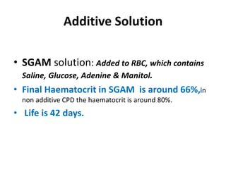
- 10. Additive Solution • SGAM solution: Added to RBC, which contains Saline, Glucose, Adenine & Manitol. • Final Haematocrit in SGAM is around 66%,in non additive CPD the haematocrit is around 80%. • Life is 42 days.
- 11. SAGM • SAGM is currently the standard additive solution used in blood banking infrastructures in the EU. • Glucose and Adenine serve to maintain adenosine triphosphate (ATP) and 2,3- diphosphoglycerate (2,3-DPG) levels, while Mannitol has functions in maintaining membrane integrity and combating hemolysis.
- 12. Storing of Blood before component Preparation Whole blood (WB) can be held at room temperature (18-25°C) up to 8 hours after collection; thereafter the unit must be refrigerated. Refrigerating it makes it unsuitable for platelet (PLT) production. Storage of whole blood at room temperature for 72 hours leads to marked reductions in pH and DPG, but the reduction in PLT function and plasma coagulation factor activity modest.
- 13. Preparation of Red Cells Red blood cells (packed red cells) are prepared by removing most of the plasma from a unit of whole blood. • Red cells have higher specific gravity than plasma, the red cells settle in the lower portion of bag due to the gravitational settling (sedimentation) or centrifugation. • The plasma is transferred into a satellite bag. • Lastly the additive solution is added to red cells.
- 14. (Sedimentation ) • The blood after collection is kept upright in refrigerator at 2-6°C, the red cells settle down and the clear supernatant plasma is transferred into a satellite bag.
- 15. Red Cells Preparation by Centrifugation • 1. Collect the appropriate volume of donor blood in primary bag of additive system, consisting of a primary bag containing anticoagulant solution CPD or CP2D attached with at least two satellite bags, one of which is empty and another contains 100 ml of additive solution e.g. Adsol or SAGM. • 2. Store at 2-6oC till processed. • 3. Centrifuge at heavy spin as above. • 4. Remove most of the supernatant plasma in the empty satellite bag. • 5. Add the additive solution to the red cells. • 6. Keep the red cells at 2-6°C and plasma at -30°C or below.
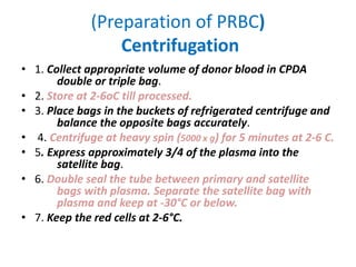
- 16. (Preparation of PRBC) Centrifugation • 1. Collect appropriate volume of donor blood in CPDA double or triple bag. • 2. Store at 2-6oC till processed. • 3. Place bags in the buckets of refrigerated centrifuge and balance the opposite bags accurately. • 4. Centrifuge at heavy spin (5000 x g) for 5 minutes at 2-6 C. • 5. Express approximately 3/4 of the plasma into the satellite bag. • 6. Double seal the tube between primary and satellite bags with plasma. Separate the satellite bag with plasma and keep at -30°C or below. • 7. Keep the red cells at 2-6°C.
- 17. Whole Blood Centrifuge RBC+ Buffy coat+ Plasma. Whole Blood Filtration - Plasma + RBC
- 18. Haematocrit & Content of different types of cells in Red Cells preparations : • A.)- Sedimented red cells: They have a PCV of 60-70 percent, 30 per cent of plasma and all original Leucocytes and platelets. • B.)- Centrifuged red cells: They have a PCV of 70- 80 percent, 10-20 percent of plasma and all original Leucocytes and platelets, • C.)- Red cells with additive (Adsol or SAG-M): They have PCV of 50-60 percent, minimum plasma and all Leucocytes and platelets.
- 19. Preparation of Platelets from Whole Blood • Platelets can be prepared either from PRP of from Buffy coat.
- 21. BUFFY COAT METHOD Compared to the PRP preparation method, the first step in buffy coat method is high g-force centrifugation followed by a low g-force step. • A buffy coat layer is obtained between the red cells and the plasma which appear after primary centrifugation. • The red cell and the plasma are transferred to their respective bags and sealed. • The satellite bag containing the buffy coat along with some plasma and red cells is the hanged and allowed to rest. Followed by a soft spin centrifugation, the supernatant platelet is separated and drawn off in a platelet bag and the remaining buffy coat is discarded after separation. • The buffy coat method yields more plasma, greater red cell loss, better initial white blood cell (WBC) reduction before filtration, and moderate reduction in viable bacteria in the platelets.
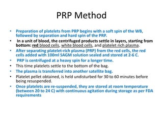
- 22. PRP Method • Preparation of platelets from PRP begins with a soft spin of the WB, followed by separation and hard spin of the PRP. • In a unit of blood, the centrifuged products settle in layers, starting from bottom: red blood cells, white blood cells, and platelet rich plasma. • After separating platelet-rich plasma (PRP) from the red cells, the red cells added with 100ml SAGM solution sealed and stored at 2-6 C. • PRP is centrifuged at a heavy spin for a longer time. • This time platelets settle to the bottom of the bag. • The plasma is transferred into another satellite bag. • Platelet pellet obtained, is held undisturbed for 30 to 60 minutes before being resuspended. • Once platelets are re-suspended, they are stored at room temperature (between 20 to 24 C) with continuous agitation during storage as per FDA requirements
- 23. Step -1, Preparation of PRP 1. Whole blood units intended for platelet preparation were placed in diagonally opposite cups of refrigerated centrifuge and temperature set at 20–24°C . 2. Then these blood bags centrifuged, - light spin (1300 rpm for 13 minutes). 3. The supernatant PRP was transferred to a satellite bag and red cells transferred the optimal additive solution (saline, adenine, glucose, mannitol; SAG-M) to the primary bag containing red cells. A dielectric sealer was used to seal the tubing between the primary bag and the satellite bag. 4. After proper labeling, the separate primary bag containing the red cells was then stored at 2–6°C. 5 . Then the PRP was processed to yield concentrate of platelets.
- 24. Step 2: Preparation of platelet concentrate from PRP • Bags containing PRP were then centrifuged for 15 minutes at 20–24°C at 3000 rpm (heavy spin). • Supernatant platelet poor plasma (PPP) was transferred to the second satellite bag, leaving about 40–70 mL of plasma for concentrated platelet resuspension. • Bag containing platelet concentrate was left undisturbed for 1–2 hours. At room temperature in an air-conditioned room. • At the end of one hour, the platelets in the resident autologous plasma were evenly resuspended by hand manipulating the platelet storage bags. • The Platelets are stored at room temperature in Agitators. • FFP is labeled and then stored at -30°C or below.
- 25. Separation of Platelet from WB. Whole Blood • Whole Blood at room temperature ( 22-34 C) soft spin (1300 ) for 13 minutes , the RBCs settled to bottom, plasma is separated using plasma extractor, Packed red cells transferred to a bag containing SAGM solution 100ml. Platelet rich Plasma • Heavy spin ( 3000 G) room temperature 22-24 C) for 15 minutes. Platelets concentrate • Platelets sinks to bottom, supernatant plasma is collected in a separate bag and frozen to -30C. • Platelets collected in a separate bag.
- 26. How Platelets are stored ? Platelet incubators at 22-24 degree C.
- 27. Platelets Platelets for transfusion can be stored five days at +22 °C, the day of collection calculated as 0. At the Blood Centre, the concentrates are kept in a special cabinet with moving shelves, which ensures that the platelets remain viable and at full value.
- 28. Precautions in Platelet Preparation The flow of blood should be uninterrupted and continuous. (If any unit takes more than eight minutes to draw, it is not suitable for preparation of platelet concentrate, fresh frozen plasma or cryoprecipitate). • Monitor the collection of blood with Automatic Mixer/Scale which is used for collecting the desired amount of blood and mixing the blood with the anticoagulant. • If platelets are to be harvested, the blood bag should be kept at room temperature (20-24°C)until platelets are separated. • Platelets should be separated within 6-8 hours from the time of collection of blood.
- 29. Quality testing of platelet concentrates . The quality of platelets during storage can be evaluated by determining the recovery and survival of the transfused platelets in thrombocytopaenic patients. A 1 h post-transfusion CCI of 10–20,000 is considered a good response while a CCI of less than 7500 is a poor response.
- 30. Quality assessment of platelet concentrates ( Whole Blood) • 1. Total platelet count of the unit.(Total number of platelets/unit of platelet concentrate = platelet count/μL of the platelet concentrate × 103 × volume of residual plasma present in the unit.) • 2. Measurement of pH of platelet concentrate unit.(This method of pH measurement permitted the differentiation of pH values within the range of 5–9.) • 3. Measurement of volume of platelet concentrate unit.(The volume was indirectly calculated by weighing the blood bag using a digital weighing scale.) • 4. Visual inspection of platelet concentrate unit. • 5. Residual leukocyte and RBC contamination of platelet concentrates
- 31. Platelet Preparation storage & Transport 1. Platelets should be prepared within 8 hrs of phlebotomy. 2. Whole blood for separation of platelets should be kept at 20-24 degree C before processing. ( Lower temperatures affect platelet function) 3. Platelet can be prepared by manual as well as using automated methods . 4. Stored at 20-24 degree C in platelet agitator cum incubator to maintain platelet function. 5. Platelets should be transported in a blood transport box that keeps the temperature in the correct range of about 20 – 24 degree C. Platelets should never be REFRIGERATED and should be transfused as soon as possible.
- 32. Summery of Platelet transfusion • Preparation for platelet transfusion starts from the production of quality-approved platelet concentrates (PC) in the blood banks. • PC can be prepared from whole blood or by apheresis. • 6 whole blood unit-derived platelets equal one apheresis platelet, which contains 5X10^10 platelets per unit. • The shelf life of a PC is five days, within which it must be used. • The normal dose of platelet transfused is calculated as 10 to 15 ml/kg of the patient. • A transfusion rate of 2 to 5 ml/min is used, thereby completing the transfusion in 1 to 2 hours. Slower flow rates are used in patients at risk of fluid overload.
- 33. Q: What is Cryoprecipitate? Ans: It is the cold insoluble portion of plasma remaining after FFP has been thawed. It contains Factor VIII ,Von Willebrand factor, factor XIII and fibrinogen. • The volume of cryoprecipitate obtained is 25-30 ml. • Self life is one year.
- 34. How Cryoprecipitate is prepared? Preparation of cryoprecipitate: 1. Plasma separated from a unit of blood within 6-8 hrs of donation and rapidly frozen within 30 minutes to -30 degree C. 2. The frozen plasma is then thawed slowly at below+ 4 degree C to obtain maximum yield of cryoprecipitate . 3. Cryo is the insoluble portion, or precipitate, that remains when the liquid portion of the plasma drains away. 4. Finally is stored at -30 degree C or colder . 5.Shelf life of one year..
- 35. Steps for Preparation of Cryo 1.Whole blood is separated to Plasma & Red cells soon after collection. 2.Gently hang the bag containing the plasma into the freezing solution ensuring that the whole bag is immersed and that the attached satellite bag is placed on the side panels of the bath. Leave in the solution for ten minutes. 3.Remove the frozen plasma, and immediately commence thawing. ( Place frozen bag in a thermostatically controlled water bath at 3° C until solution becomes “slushy”); (approximately 20 - 30 minutes). 4.Immediately spin in a refrigerated centrifuge for 10 minutes at 2000 rpm at 2°- 8° C. Optimum temperature is 3° C. 5.Remove the excess plasma into the empty satellite bag leaving approximately 10 ml with the deposited cryoprecipitate. 6.Detach the excess plasma bag from the cryoprecipitate.
- 36. How Frozen plasma and cryoprecipitate is transported? During transport, frozen components must be maintained at or below the required storage temperature. This can be achieved with a suitable quantity of wet ice or dry ice ( dry ice temperature = minus 78.5 0 Celsius) in well insulated containers or standard shipping cartons lined with insulating material such as plastic air bubble packaging or dry packaging fragments.
- 37. Flow Chart for Preparation of Cryoprecipitate 1 •Bleed Donor 2 •Centrifuge ( 3000 RPM for 20 minutes) 3 •Separate Plasma and Red Cells 4 •Snap Fridge ( Dry Ice & alcohol -60 c for 10 minutes) 5 •Thawing till Slushy ( 3 c for 20-30minutes) 6 •Quantity Released ( Stored at -20 c or Lower)
- 38. FFP • Fresh Frozen Plasma (FFP) is the liquid part of the blood obtained after separation of the cellular part of the blood. • FFPs have a volume of 200–300 mL, • Separated liquid part is immediately frozen, to a temperature of –25° C . • FFPs primarily contain stable clotting factors, albumin and Immunoglobulins. • It should be prepared and stored within 6 hours, to ensure best activity of the coagulation factors. • FFPs can be stored for up to one year at a temperature of –25° C. • However, prior to administration, FFPs would be needed to be thawed, and need to be used within 30 minutes of thawing. • Generally, the dose of FFPs ranges 10–15 mL/kg. • The target INR is 1.7, prothrombin time (PT) a activated Prothrombin time (APTT) of less than twice the normal.
- 39. Equipment 1. Refrigerated blood bank centrifuge 2. Blood bank refrigerator 3. Freezer –20º C or below 4. Platelet agitator 5. Weighing scale for balancing centrifuge cups 6. Tube sealer 7. Plasma Defroster or Water Bath 8. Plasma expresser (manual) 9. Component Separator 10. Stripper/ Cutter 11. Aluminum canisters for cryoprecipitate
- 40. Consumable items • 1. Sealing rings, Rubber Bands • 2. Plastic boxes for storing fresh frozen plasma in freezer • 3. Double, Triple and quadruple blood bags • 4. Appropriate sticker type labels and instructions for each component • 5. Gloves
Editor's Notes
- #3: Red cells ( RBC), WBC, Platelets, Plasma.
- #6: All current methods for separation and preparation of the three major blood components— RBCs, platelets, and plasma—rely on one or more centrifugation steps
- #30: Calculation of corrected count increment (CCI) : corrects for the patient’s blood volume and the number of platelets in the PC when evaluating the post-transfusion platelet count increment.
- #32: There was a gradual loss of platelet discoid shape at exposure temperatures < 20 degrees C, which worsened as temperatures decreased and exposure times increased to 24 hours.
- #36: 7. If storage is at -20° C, the expiry period is 6 months. 8. If storage is at -40° C, the expiry period is 12 months