Downloaded 179 times


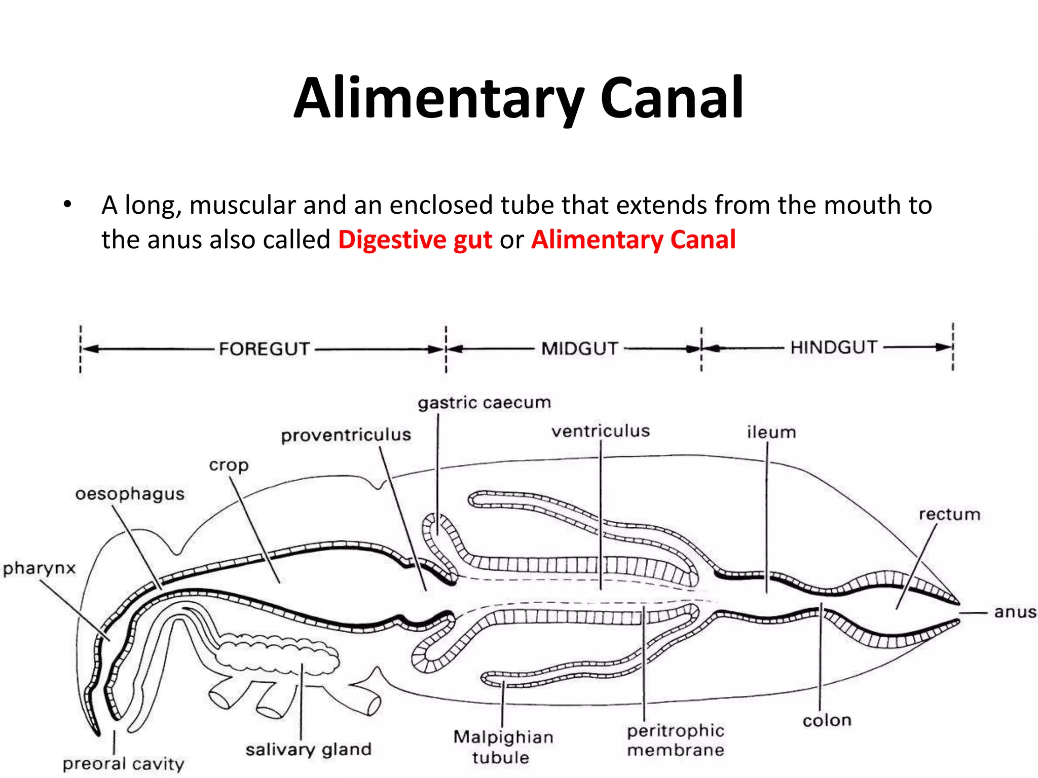


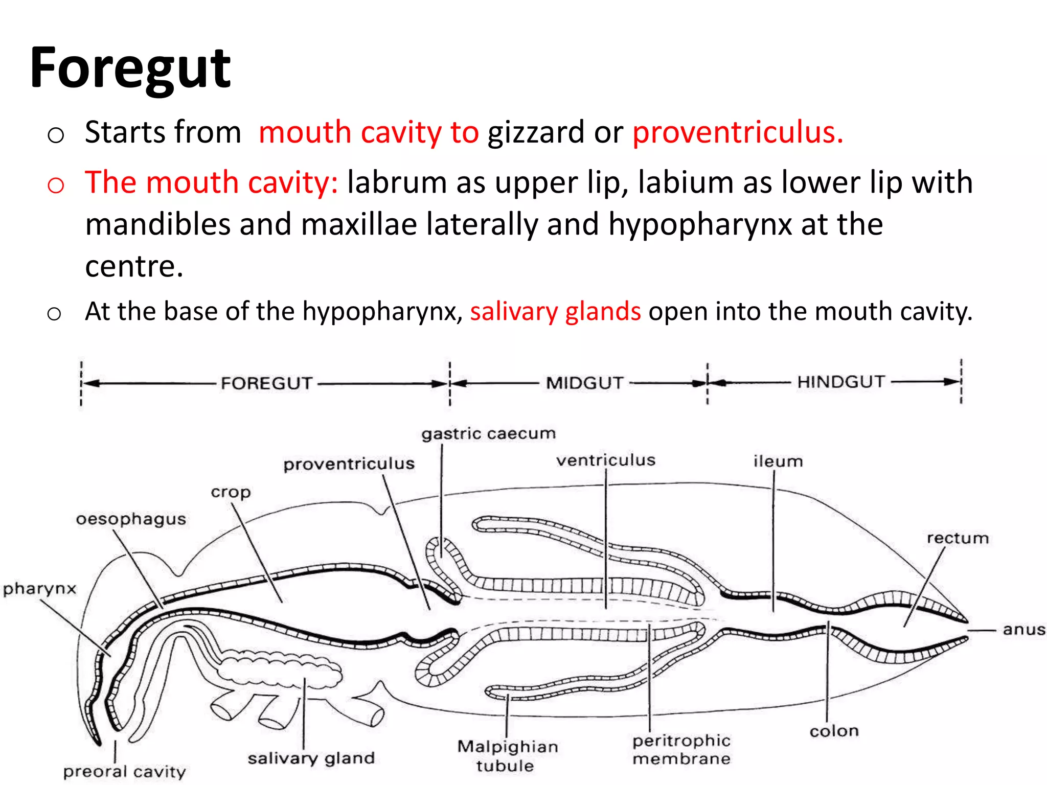


The document summarizes the digestive system of insects. It describes that insects have a long alimentary canal divided into foregut, midgut, and hindgut. The foregut contains the mouth, pharynx, esophagus, crop, and gizzard. Salivary glands secrete enzymes into the foregut. The midgut is the primary site of digestion and absorption, containing gastric caeca that secrete enzymes. The hindgut absorbs nutrients and forms feces. Digestion involves ingestion, transportation, digestion in the midgut by enzymes, absorption of nutrients, and egestion of waste.


