Approach to HEART FAILURE Dr. Subroto Mandal, MD, DM Assistant Professor, Cardiology NRI Heart Centre & Research Institute
Definition of Heart Failure HF is a complex clinical syndrome that can result from any structural or functional cardiac disorder that impairs the ability of the ventricle to fill with or eject blood. ACC/AHA Task force
“ Heart Failure” vs. “Congestive Heart Failure” Because not all patients have volume overload at the time of initial or subsequent evaluation, the term “heart failure” is preferred over the older  term “congestive heart failure.”
Relatively common disorder The incidence of HF approaches 10 per 1000 population after age 65 1-2 % at the age of 45-50yrs >10% at the age >75yrs At 40yrs age life time risk for HF 21%for men 20.3% for women 80% admission for HF > 65yrs old Cost of hospitalization for heart failure is twice that for all form of cancer and myocardial infarction combined PREVALENCE & INCIDENCE
CLASSIFICATION Forward Vs Backward Rt. Vs Lt. sided HF  Acute Vs Chronic HF Low Vs High output HF  Systolic Vs Diastolic HF
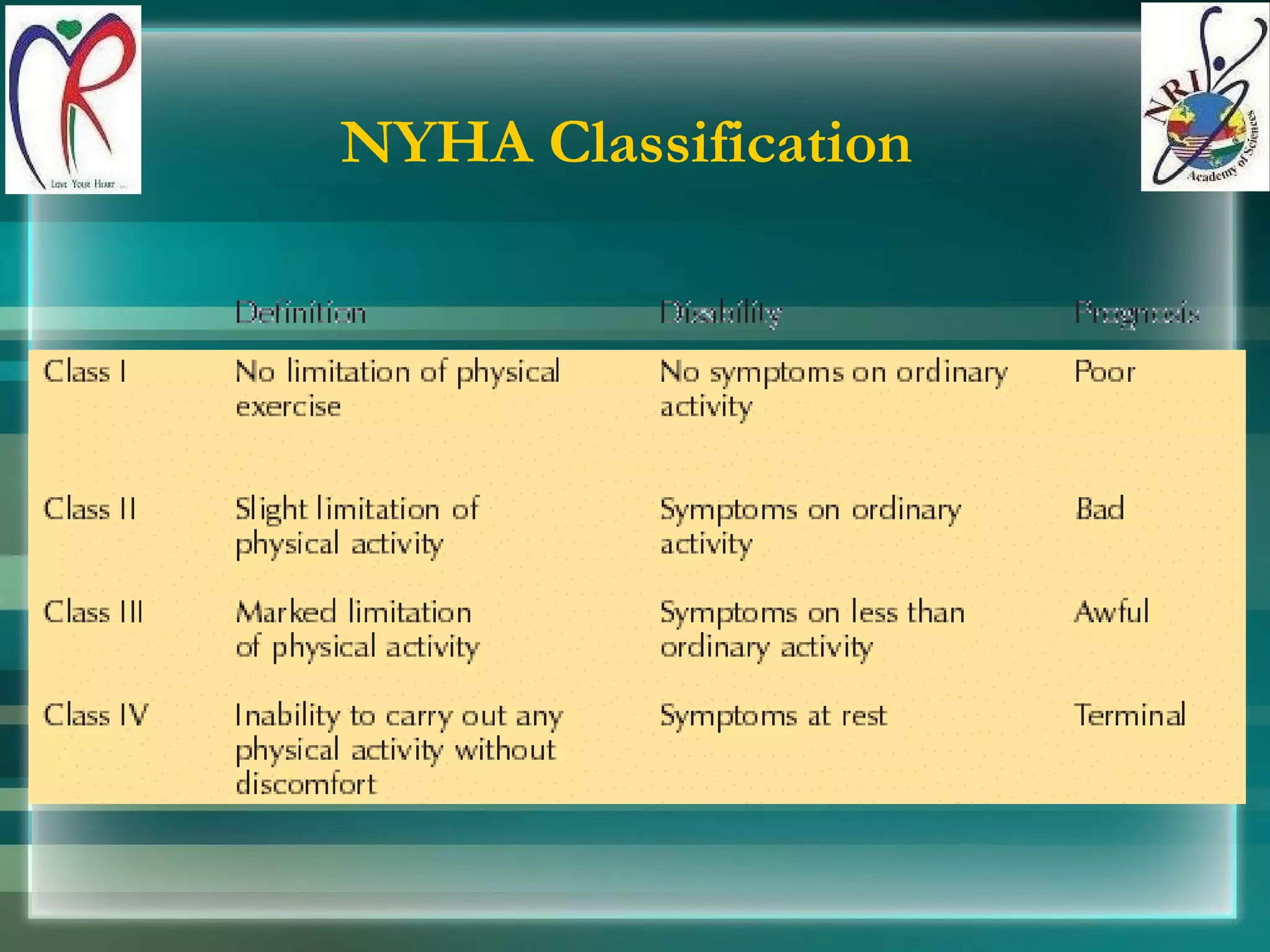
NYHA Classification
Stages of Heart Failure At Risk for Heart Failure: STAGE A   High risk for developing HF STAGE B   Asymptomatic LV dysfunction Heart Failure: STAGE C   Past or current symptoms of HF STAGE D   End-stage HF
Stages of Heart Failure Designed to emphasize  preventability  of HF Designed to recognize the  progressive nature  of LV dysfunction
Stages of Heart Failure COMPLEMENT, DO  NOT  REPLACE NYHA CLASSES NYHA Classes - shift back/forth in individual patient  (in response to Rx and/or progression of disease) Stages - progress in  one  direction due to cardiac remodeling
 
Rapid classification of hemodynamic states
PRECIPITATING FACTORS INAPPROPRIATE  THERAPY HIGH SALT INTAKE  ARRYTHMIAS INFARCTION OR ISCHAEMIA PULMONARY EMBOLISM SYSTEMIC INFECTION PHYSICAL & EMOTIONAL STRESS INFECTIVE ENDOCARDITIS COMORBIDITY ( renal failure, sepsis) MYOCARDIAL DEPRESSANT DRUGS CARDIAC TOXINS HIGH OUTPUT STATES
 
Evaluation of HF patient
 
 
RIGHT SIDED SYMPTOMS Abdominal Pain Nausea Constipation Anorexia Bloating Ascites SIGNS Peripheral edema Hepatomegaly JVD or JVP HJR LEFT SIDED DOE PND Orthopnea Tachypnea Cough Hemoptysis Bibasilar rales Pulmonary edema S3 gallop Pleural effusion Cheyne-Stokes respiration
COUGH Caused by pulmonary congestion Nonproductive cough in LVF (Dyspnea equivalent) Cough at recumbency (Orthopnea equivalent)
JVP Normal upper limit of JVP are 4 cm from sternal angle TR  V wave  and Y descend are prominent Kussumal sign  in constrictive pericarditis
 
Right heart failure
INVESTIGATIONS BNP ECG CXR ECHOCARDIOGRAPHY ( TTE, TEE) CARDIAC CATHETERIZATION MUGA SCAN, CT, MRI ANGIO ENDOMYOCARDIAL BIOPSY VIABILITY ASSESMENT (DSE, MRI, SPECT, PET scan) ARRYTHMIA WORK UP
Atrial fibrillation
Myocarditis
Cardiomegaly
Dextrocardia – Acute pulmonary edema
CCF
Mitral stenosis
Pericardial effusion
Mitral Stenosis
Mitral Stenosis Pre PBMV Post PBMV
MVP - MR
Aortic Stenosis
LA Myxoma
Post MI-VSD
DCM
DCM
DCM
MR Coronary Angiogram
CT Coronary Angiogram
HCM
Framingham Criteria for Congestive Heart Failure Major criteria:           Paroxysmal nocturnal dyspnea           Neck vein distention          Rales          Radiographic cardiomegaly     Acute pulmonary edema          S3 gallop          Increased central venous pressure (>16 cm H2O at right atrium)          Hepatojugular reflux          Weight loss  >4.5 kg in 5 days in response to treatment   Minor criteria:            Bilateral ankle edema          Nocturnal cough          Dyspnea on ordinary exertion          Hepatomegaly          Pleural effusion          Decrease in vital capacity by one third from maximum recorded          Tachycardia (heart rate>120 beats/min.)
 

Heart failure

  • 1.
    Approach to HEARTFAILURE Dr. Subroto Mandal, MD, DM Assistant Professor, Cardiology NRI Heart Centre & Research Institute
  • 2.
    Definition of HeartFailure HF is a complex clinical syndrome that can result from any structural or functional cardiac disorder that impairs the ability of the ventricle to fill with or eject blood. ACC/AHA Task force
  • 3.
    “ Heart Failure”vs. “Congestive Heart Failure” Because not all patients have volume overload at the time of initial or subsequent evaluation, the term “heart failure” is preferred over the older term “congestive heart failure.”
  • 4.
    Relatively common disorderThe incidence of HF approaches 10 per 1000 population after age 65 1-2 % at the age of 45-50yrs >10% at the age >75yrs At 40yrs age life time risk for HF 21%for men 20.3% for women 80% admission for HF > 65yrs old Cost of hospitalization for heart failure is twice that for all form of cancer and myocardial infarction combined PREVALENCE & INCIDENCE
  • 5.
    CLASSIFICATION Forward VsBackward Rt. Vs Lt. sided HF Acute Vs Chronic HF Low Vs High output HF Systolic Vs Diastolic HF
  • 6.
  • 7.
    Stages of HeartFailure At Risk for Heart Failure: STAGE A High risk for developing HF STAGE B Asymptomatic LV dysfunction Heart Failure: STAGE C Past or current symptoms of HF STAGE D End-stage HF
  • 8.
    Stages of HeartFailure Designed to emphasize preventability of HF Designed to recognize the progressive nature of LV dysfunction
  • 9.
    Stages of HeartFailure COMPLEMENT, DO NOT REPLACE NYHA CLASSES NYHA Classes - shift back/forth in individual patient (in response to Rx and/or progression of disease) Stages - progress in one direction due to cardiac remodeling
  • 10.
  • 11.
    Rapid classification ofhemodynamic states
  • 12.
    PRECIPITATING FACTORS INAPPROPRIATE THERAPY HIGH SALT INTAKE ARRYTHMIAS INFARCTION OR ISCHAEMIA PULMONARY EMBOLISM SYSTEMIC INFECTION PHYSICAL & EMOTIONAL STRESS INFECTIVE ENDOCARDITIS COMORBIDITY ( renal failure, sepsis) MYOCARDIAL DEPRESSANT DRUGS CARDIAC TOXINS HIGH OUTPUT STATES
  • 13.
  • 14.
  • 15.
  • 16.
  • 17.
    RIGHT SIDED SYMPTOMSAbdominal Pain Nausea Constipation Anorexia Bloating Ascites SIGNS Peripheral edema Hepatomegaly JVD or JVP HJR LEFT SIDED DOE PND Orthopnea Tachypnea Cough Hemoptysis Bibasilar rales Pulmonary edema S3 gallop Pleural effusion Cheyne-Stokes respiration
  • 18.
    COUGH Caused bypulmonary congestion Nonproductive cough in LVF (Dyspnea equivalent) Cough at recumbency (Orthopnea equivalent)
  • 19.
    JVP Normal upperlimit of JVP are 4 cm from sternal angle TR V wave and Y descend are prominent Kussumal sign in constrictive pericarditis
  • 20.
  • 21.
  • 22.
    INVESTIGATIONS BNP ECGCXR ECHOCARDIOGRAPHY ( TTE, TEE) CARDIAC CATHETERIZATION MUGA SCAN, CT, MRI ANGIO ENDOMYOCARDIAL BIOPSY VIABILITY ASSESMENT (DSE, MRI, SPECT, PET scan) ARRYTHMIA WORK UP
  • 23.
  • 24.
  • 25.
  • 26.
    Dextrocardia – Acutepulmonary edema
  • 27.
  • 28.
  • 29.
  • 30.
  • 31.
    Mitral Stenosis PrePBMV Post PBMV
  • 32.
  • 33.
  • 34.
  • 35.
  • 36.
  • 37.
  • 38.
  • 39.
  • 40.
  • 41.
  • 42.
    Framingham Criteria forCongestive Heart Failure Major criteria:        Paroxysmal nocturnal dyspnea         Neck vein distention         Rales         Radiographic cardiomegaly    Acute pulmonary edema         S3 gallop         Increased central venous pressure (>16 cm H2O at right atrium)         Hepatojugular reflux         Weight loss >4.5 kg in 5 days in response to treatment   Minor criteria:         Bilateral ankle edema         Nocturnal cough         Dyspnea on ordinary exertion         Hepatomegaly         Pleural effusion         Decrease in vital capacity by one third from maximum recorded         Tachycardia (heart rate>120 beats/min.)
  • 43.

Editor's Notes

  • #12 Figure 7-17. Rapid classification of hemodynamic states. Stevenson [42] popularized the concept of tailoring therapies to the hemodynamic status of patients. This approach can be coupled to a noninvasive diagnostic evaluation of patients admitted to the hospital with congestive heart failure with therapeutic approaches, perhaps directed by varying combinations of fluid retention states and peripheral organ perfusion. The two basic components outlined in this figure are, indeed, congestion (B and C) and low perfusion (C and D). Perhaps the most complex patient is the individual who is substantially volume overloaded with low flow states (D). These patients as well as those with simply low perfusion states are generally in cardiogenic shock. Signs and symptoms of congestion to review include orthopnea, paroxysmal nocturnal dyspnea, jugular venous distention, hepatomegaly, particularly with hepatojugular reflux, peripheral edema, presence of rales (remembering that they can be rare in chronic heart failure), and the Valsalva square wave blood pressure sign. Signs of low perfusion include a narrow pulse pressure, a sleepy or obtunded patient, periodic respirations, cool extremities, hypotension after angiotensin-converting enzyme inhibitor introduction, and renal dysfunction or a low serum sodium [42].
  • #15 Figure 7-1. Specific goals of patient evaluation when heart failure is suspected. First, one must appropriately recognize the heart failure syndrome and differentiate heart and circulatory failure from problems that cause similar complaints and findings. Second, by staging the severity of heart failure, the clinician can establish prognosis with reasonable accuracy. This is important in the design of therapeutic protocols to treat certain aspects of the syndrome. Finally, identifying the primary etiology of myocardial dysfunction and determining the precipitating causes of decompensation are extremely important. The interplay of patient history, physical examination, laboratory tests, and specific diagnostic studies helps the clinician achieve these goals.