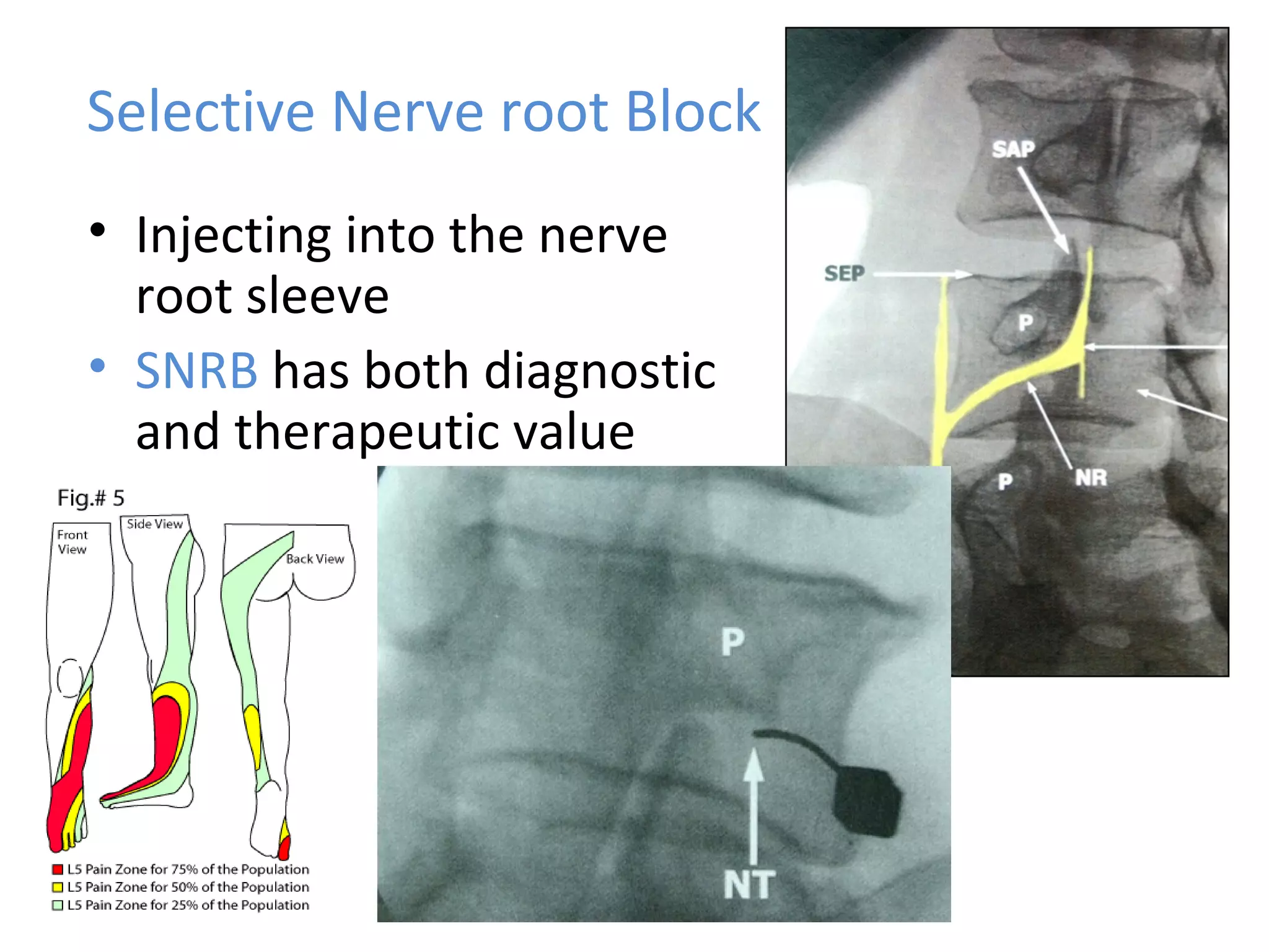
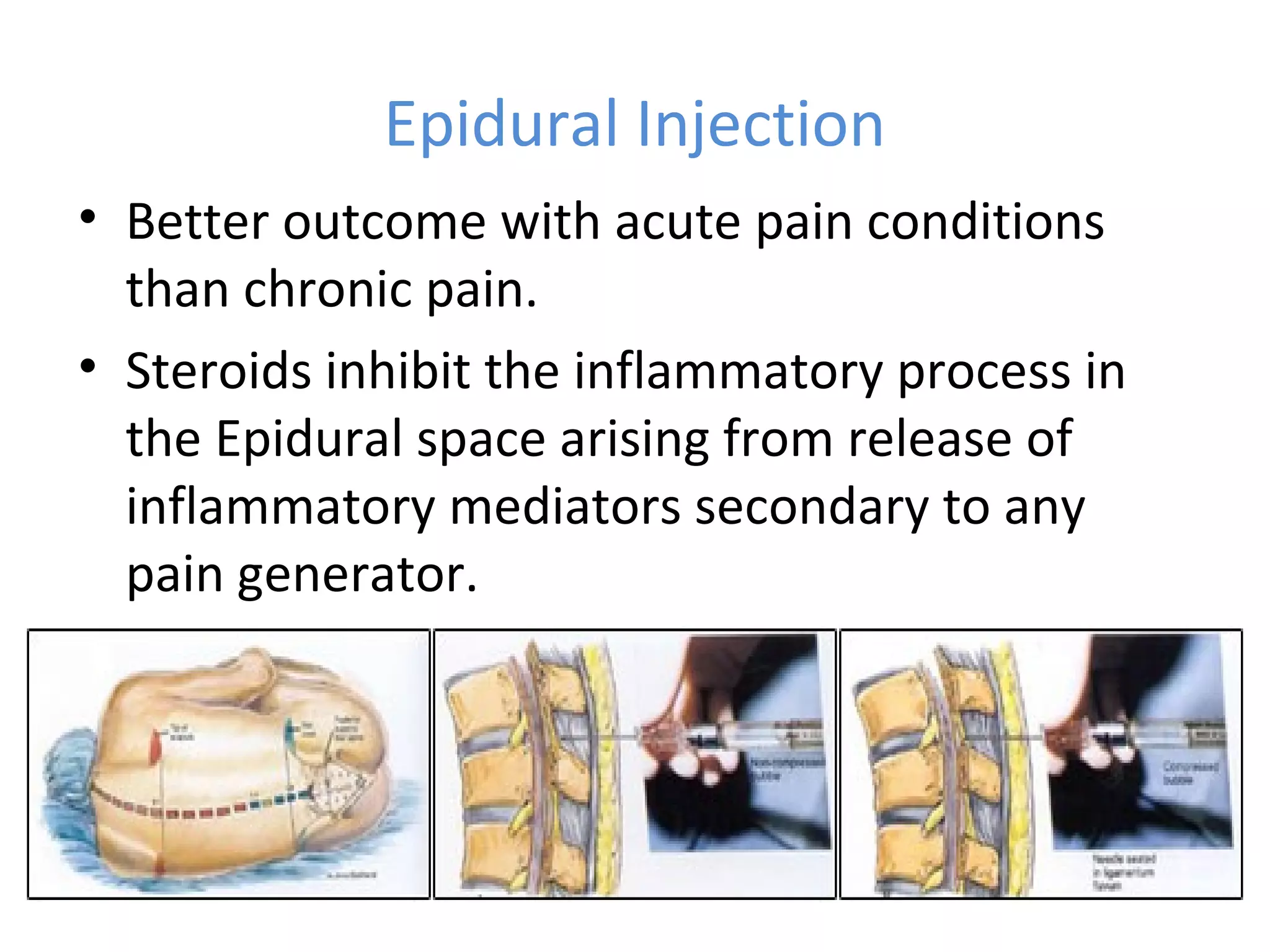
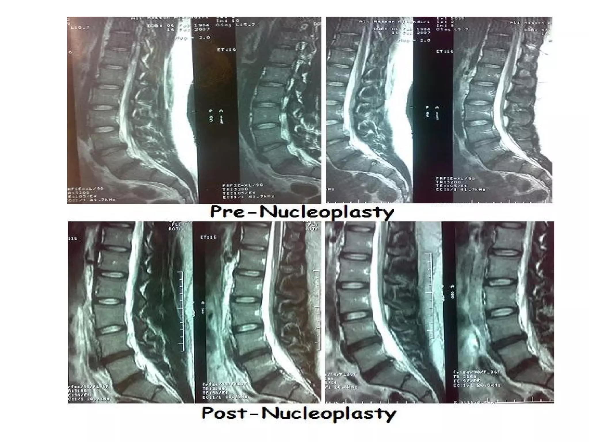
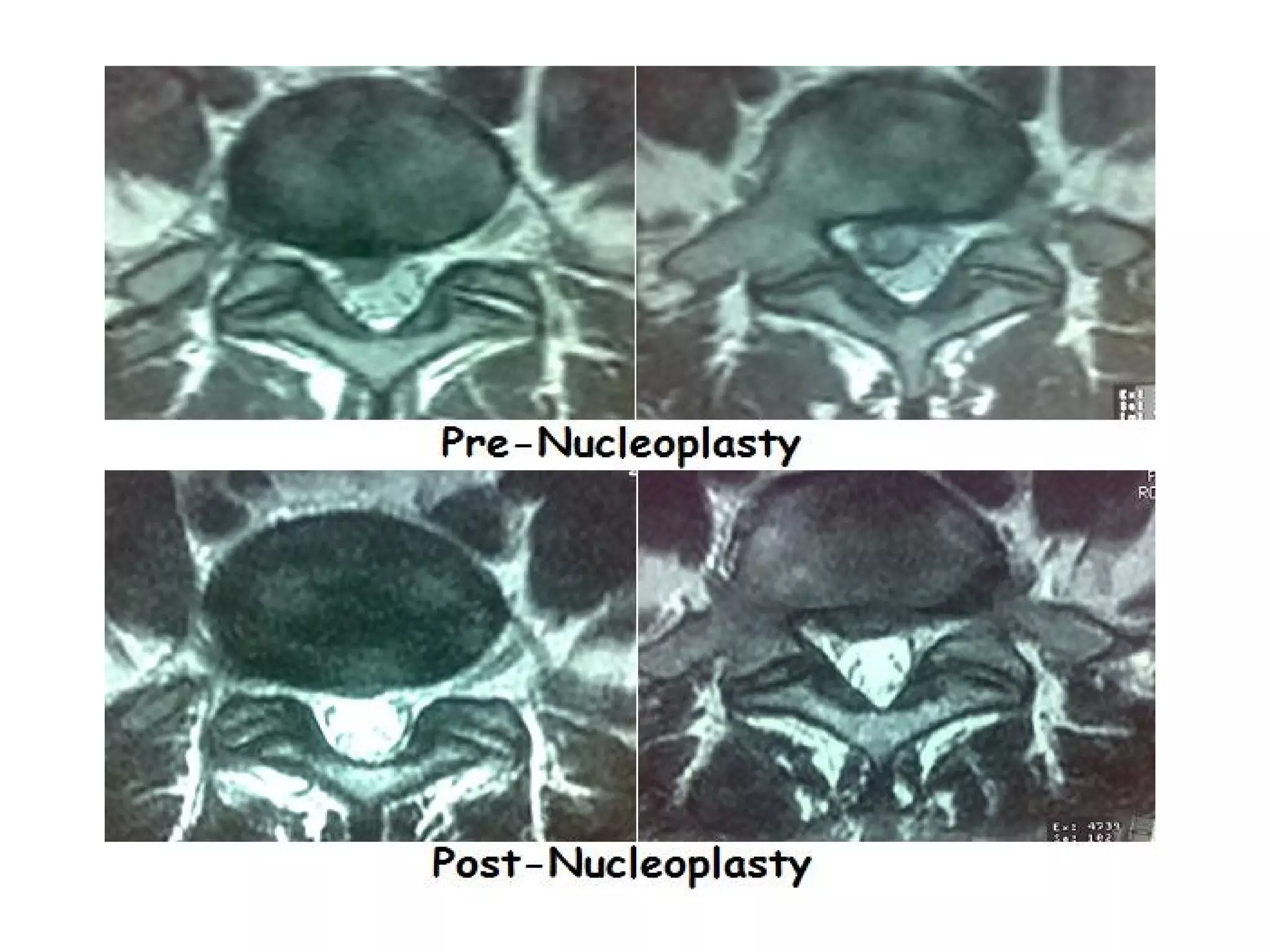
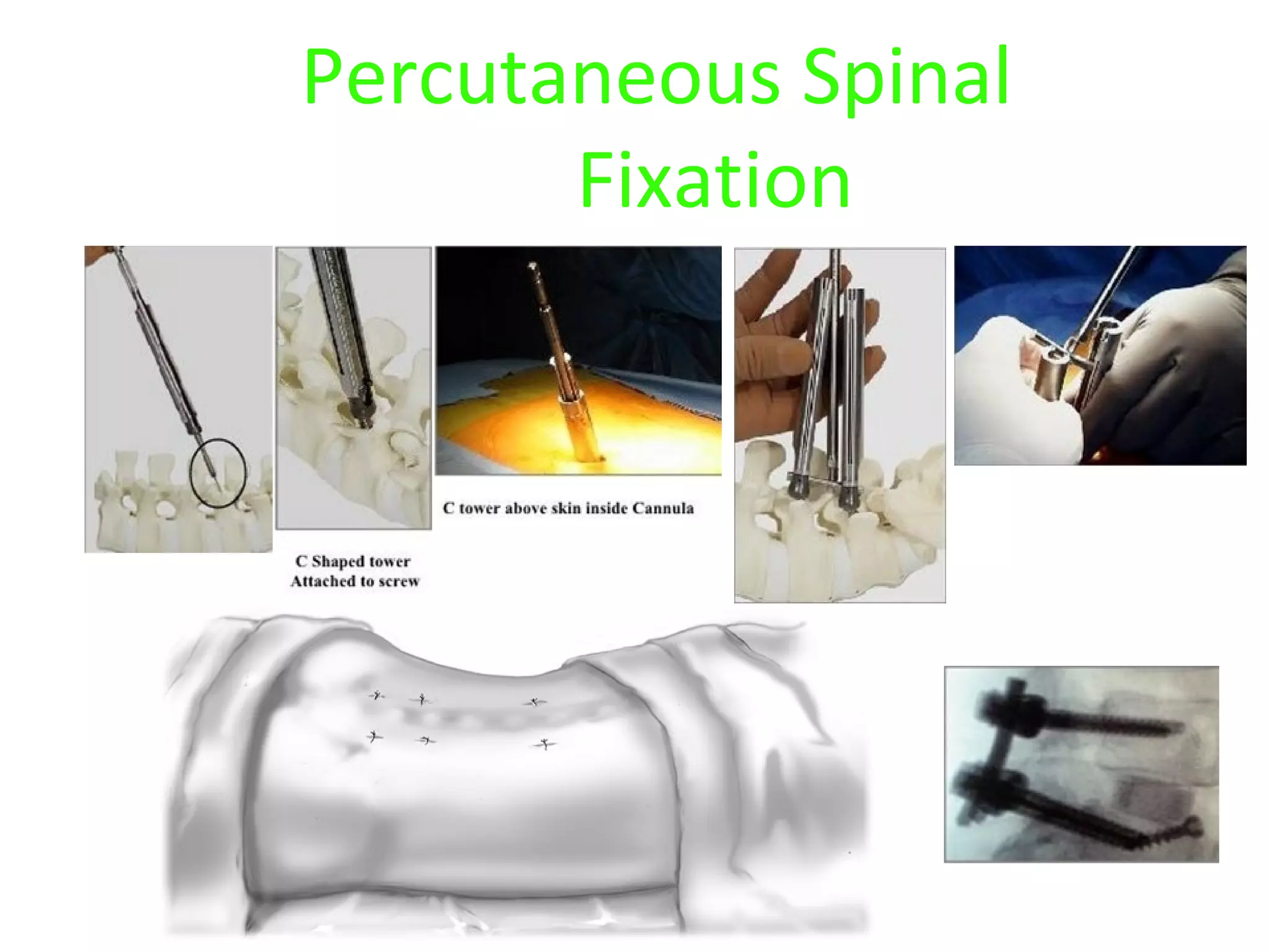
Minimally Invasive Techniques (MIT) in
Management of Lumbar Disc Disease
(New Trends in Spine Interventions)
Mohamed Mohi Eldin
Prof. of Neurosurgery,
Faculty of Medicine,
Cairo University
Less is more
Causes of Low back Pain
• Muscle Spasm
• Disc herniation (bulge, protrusion,
extrusion)
• Annulus Fissures
• Facet disease
• Spinal Canal stenosis
• Saco-iliac disease
• Spondylolisthesis
• V.C.F. (Osteoprosis, Tumours)
• Biopyschosocial model
Back pain
• Treating back pain is
a big problem.
• 80% of all people will experience
back pain in their life
• Traditionally people fear back pain
(? Paralysis )
• No proper consensus on best
treatment.
History taking is the most
important part of
Diagnosing the condition
Changing the look to MRI
( how many MRIs formerly
seen as –ve ?)
Discogenic Pain
Facetogenic Pain
Foraminal Pain
Conservative treatment still
first-line (8-10 Ws)
Injections
Nucleoplasty
Endoscopic Approaches
Percutaneous Techniques
Microsurgical Techniques
Open Surgery
M
O
R
E
I
N
V
A
S
I
V
E
More
Improvements
Less
Invasiveness
Definition of MIT
• Treatment that requires no or
minimal dissection or cutting.
• Under Local anaesthesia, mild
sedation or epidural.
• Day case or one day
hospitalisation.
• Quick recovery and results.
• Side effects are minimal or none.
• Usually under fluoroscopy.
Role of Minimally Invasive
Interventions
Many Minimally-Invasive Methods :
1. Discography
2. Epidural Injection
3. Facet / median nerve block and Ablation.
4. Selective Nerve root block
5. Sacro-iliac joint injection
6. Nucleoplasty
7. Vertebroplasty and Kyphoplasty
8. Cavity assisted Tumour Debulking
9. Percutaneous Intraspinous spacer
10. Percutaneous Spinal Fixation
General Contra-Indications to MIT
• Conditions that require surgery
• Progressive or Neurological Deficit
• Evidence of Infection
• Coagulopathy
• Other disease causing the pain
• Pregnancy.
Discography
• Assessing disc morphology + pain
response
• More sensitive than MRI in detecting
Fissure as a cause of LBP
• 5 grades. Modified Dallas
Classification
Facet joint & Median branch block
• Diagnostic and
Therapeutic
• Intra or
Extra- articular
Selective Nerve root Block
• Injecting into the nerve
root sleeve
• SNRB has both diagnostic
and therapeutic value
Epidural Injection
• Better outcome with acute pain conditions
than chronic pain.
• Steroids inhibit the inflammatory process in
the Epidural space arising from release of
inflammatory mediators secondary to any
pain generator.
Indications
Interlaminar Epidural
• Spinal canal compromise
• Disc Degeneration or
herniation
Transforaminal Epidural
• Spinal root compression.
• Spinal root Inflammation.
Sacroiliac Joint Injection
• Another cause of LBP
• Degenerative or Inflammatory
• Difficult to diagnose Clinically
• S-I joint injection is the best
diagnostic tool
• Plain x-ray is enough.
• Intra-articular or Extra-articular
injection.
• Image Guidance is essential for
accurate needle placement
NucleoplastyNucleoplasty
(Percutaneous disc decompression)(Percutaneous disc decompression)
• Under mild sedation and LocalUnder mild sedation and Local
• Day case.Day case.
• Lasts 20-30 minutes.Lasts 20-30 minutes.
• 17 Gauge needle17 Gauge needle
• Ablation of Nucleous withAblation of Nucleous with
coblation wand through thecoblation wand through the
needleneedle
Indications:
Radicular/Axial Symptoms:
• Leg pain or back pain
• MRI evidence of
contained posterior
lateral focal disc
protrusion
• Failed conservative
therapy or selective nerve
root block (if this is done)
• Mild or no Neurological
deficit.
Contra-Indications
• Disc height < 50%
• Severe disc degeneration
• Severe spinal stenosis
• Progressive or severe Neurological Deficit
• Disc occupying one-third or more of spinal canal
• Non-contained disc.
• Evidence of Infection or Coagulopathy
• Age < 18 and > 65
Nucleoplasty (Experience)
- Introduced Oct.
2005
- Chronic LBP,
- Failed
conservative
therapy (8 – 12
weeks)
- Contained disc
protrusion
- Lumbar discs (1-3
levels)
Balloon
Kyphoplasty
Balloon Kyphoplasty Procedure
Case 3
Case 3
95% improvement. Walk next day. No leak95% improvement. Walk next day. No leak
Cavity Spine Wand
• Has a bouquet of
active electrodes at
the tip
– This is where the
plasma is formed
– The device ablates in
a “forward motion”
• Has an ‘S’ shaped tip
to maneuver off-axis
Active Electrodes
Insulator
Return
Electrode
Cavity Tumor Procedure
1-Tumor in vertebral body
2- Insert Access Cannula
Cavity Tumor Procedure
3- Deploy cavity spinewand
4- Debulk Tissue to Create Void
5- Augment with Cement
Interspinous process-based Dynamic Stabilization
Distraction (IPD) devices
Indications
1. Stenosis:
• Central, lateral and foraminal lumbar spinal
stenosis with leg, buttock or groin pain, which can
be relieved during flexion
1. Low Back Bain:
• Facet syndrome due to facet osteoarthritis
• Degenerative spondylolisthesis up to Grade I with
hyperlordotic curve
• DDD with retrolisthetis
• Interspinous pain arising from (“Kissing Spines”)
In Lumbar Canal Stenosis
• Implants are placed between adjacent spinous
processes
• to act as physical blocks to extension (and thus lumbar
canal narrowing) on standing or walking, relieving
pressure on the nerves.
• Play a role between medical symptom control and the
more invasive procedure of laminectomy.
Principle of action
• Flexion decreases the amount of
stenosis by distracting the soft
tissues
– ligamentum flavum,
– posterior annulus and
– facet capsule
• Therefore increasing the area of
the spinal canal.
• A correctly placed spacer mimics
flexion at the implanted spinal
segment.
• This usually relieves the neurogenic
claudication which improves a
patient’s function. 
The targets of interspinous spacers
are the joints at the dorsal part of the column
not the disc, which is located far away ventrally
Some Available Spacers
Posterior approach
Percutaneous, lateral approah
X’Stop Diam Coflex Wallis
In-Space
Percutaneous Interspinous Spacer (Synthes Spine)
UndeployedUndeployed DeployedDeployed
The Diam System
(Medtronic Sofamor Danek, Memphis,Tennessee)
• a deformable damper
• made of silicone,
• with a polyester jacket
• designed with oversized wings, which
contribute to the stability and
performance of the implant
• does not burn any bridges for potential
surgeries in the future
The Coflex Titanium System
(Paradigm Spine, LLC, New York)
• Functionally dynamic
– Compressible in extension, allowing
flexion
– Increased rotational stability
– Center of rotation close to spinal
canal
• Protection of posterior elements
– Stress reduction on facet joints
– Maintenance of foraminal height
• Ease of use
– Less invasive, tissue-sparing
procedure
– Easy and precise application
Representative
Cases
Flexion Extension
Case
Case
Representative
Cases
Case
5454years:Spinal stenosis L4-L5years:Spinal stenosis L4-L5
VAS Back 5, VAS Leg 8, 50m, ODI 25/50VAS Back 5, VAS Leg 8, 50m, ODI 25/50
3m FU: VAS Back 3, Leg 3
ODI 15, Free Walking
Spinal stenosis / 63 yearsSpinal stenosis / 63 years
Case exampleCase example
DDD with segmental hyperlordosis / 41 yearsDDD with segmental hyperlordosis / 41 years
Case exampleCase example
Case example
Soft disc / 41 yearsSoft disc / 41 years
Case examples
Retrolisthesis / 53 yearsRetrolisthesis / 53 years
Case examplesCase examples
Minimal Invasive Spinal Surgery
• Allows for minimally
invasive visualization
• A series of soft-tissue
dilators create a small
tunnel through the muscles
of the back
• A tube is inserted to the
level of the spinal column
• Surgery can be guided by a
video camera with
magnifying lens and fiber-
optic light source
Mini-TLIF
(Transforaminal Lumbar Interbody Fusion)
• Used in decompression
procedures
• Versatile instruments
allow a surgeon to
approach the spine using
an open, mini-open or
minimally invasive
technique
PLIF
Posterior Lumbar Interbody Fusion
• DISC SPACE DISTRACTION
Percutaneous Spinal
Fixation
Minimal Invasive Spinal Surgery
Open
• 6-8 inch incision
• Tissues are separated
and held apart during
surgery
• Hospital stay could be
days
Minimally Invasive
• 1 inch incision
• Tissues are left intact
and split along natural
divisions
• Outpatient surgery for
some
mmohi63@yahoo.com
mohamedmohieldin.com
mohamedmohieldin2.com

Minimal invasive techniques in lumbar degenerative diseases

  • 1.
    Minimally Invasive Techniques(MIT) in Management of Lumbar Disc Disease (New Trends in Spine Interventions) Mohamed Mohi Eldin Prof. of Neurosurgery, Faculty of Medicine, Cairo University
  • 5.
  • 6.
    Causes of Lowback Pain • Muscle Spasm • Disc herniation (bulge, protrusion, extrusion) • Annulus Fissures • Facet disease • Spinal Canal stenosis • Saco-iliac disease • Spondylolisthesis • V.C.F. (Osteoprosis, Tumours) • Biopyschosocial model
  • 7.
    Back pain • Treatingback pain is a big problem. • 80% of all people will experience back pain in their life • Traditionally people fear back pain (? Paralysis ) • No proper consensus on best treatment.
  • 8.
    History taking isthe most important part of Diagnosing the condition Changing the look to MRI ( how many MRIs formerly seen as –ve ?) Discogenic Pain Facetogenic Pain Foraminal Pain
  • 9.
    Conservative treatment still first-line(8-10 Ws) Injections Nucleoplasty Endoscopic Approaches Percutaneous Techniques Microsurgical Techniques Open Surgery M O R E I N V A S I V E More Improvements Less Invasiveness
  • 10.
    Definition of MIT •Treatment that requires no or minimal dissection or cutting. • Under Local anaesthesia, mild sedation or epidural. • Day case or one day hospitalisation. • Quick recovery and results. • Side effects are minimal or none. • Usually under fluoroscopy.
  • 11.
    Role of MinimallyInvasive Interventions Many Minimally-Invasive Methods : 1. Discography 2. Epidural Injection 3. Facet / median nerve block and Ablation. 4. Selective Nerve root block 5. Sacro-iliac joint injection 6. Nucleoplasty 7. Vertebroplasty and Kyphoplasty 8. Cavity assisted Tumour Debulking 9. Percutaneous Intraspinous spacer 10. Percutaneous Spinal Fixation
  • 12.
    General Contra-Indications toMIT • Conditions that require surgery • Progressive or Neurological Deficit • Evidence of Infection • Coagulopathy • Other disease causing the pain • Pregnancy.
  • 13.
    Discography • Assessing discmorphology + pain response • More sensitive than MRI in detecting Fissure as a cause of LBP • 5 grades. Modified Dallas Classification
  • 14.
    Facet joint &Median branch block • Diagnostic and Therapeutic • Intra or Extra- articular
  • 15.
    Selective Nerve rootBlock • Injecting into the nerve root sleeve • SNRB has both diagnostic and therapeutic value
  • 16.
    Epidural Injection • Betteroutcome with acute pain conditions than chronic pain. • Steroids inhibit the inflammatory process in the Epidural space arising from release of inflammatory mediators secondary to any pain generator.
  • 17.
    Indications Interlaminar Epidural • Spinalcanal compromise • Disc Degeneration or herniation Transforaminal Epidural • Spinal root compression. • Spinal root Inflammation.
  • 19.
    Sacroiliac Joint Injection •Another cause of LBP • Degenerative or Inflammatory • Difficult to diagnose Clinically • S-I joint injection is the best diagnostic tool • Plain x-ray is enough. • Intra-articular or Extra-articular injection. • Image Guidance is essential for accurate needle placement
  • 20.
    NucleoplastyNucleoplasty (Percutaneous disc decompression)(Percutaneousdisc decompression) • Under mild sedation and LocalUnder mild sedation and Local • Day case.Day case. • Lasts 20-30 minutes.Lasts 20-30 minutes. • 17 Gauge needle17 Gauge needle • Ablation of Nucleous withAblation of Nucleous with coblation wand through thecoblation wand through the needleneedle
  • 21.
    Indications: Radicular/Axial Symptoms: • Legpain or back pain • MRI evidence of contained posterior lateral focal disc protrusion • Failed conservative therapy or selective nerve root block (if this is done) • Mild or no Neurological deficit.
  • 22.
    Contra-Indications • Disc height< 50% • Severe disc degeneration • Severe spinal stenosis • Progressive or severe Neurological Deficit • Disc occupying one-third or more of spinal canal • Non-contained disc. • Evidence of Infection or Coagulopathy • Age < 18 and > 65
  • 23.
    Nucleoplasty (Experience) - IntroducedOct. 2005 - Chronic LBP, - Failed conservative therapy (8 – 12 weeks) - Contained disc protrusion - Lumbar discs (1-3 levels)
  • 30.
  • 31.
  • 32.
  • 33.
    Case 3 95% improvement.Walk next day. No leak95% improvement. Walk next day. No leak
  • 34.
    Cavity Spine Wand •Has a bouquet of active electrodes at the tip – This is where the plasma is formed – The device ablates in a “forward motion” • Has an ‘S’ shaped tip to maneuver off-axis Active Electrodes Insulator Return Electrode
  • 35.
    Cavity Tumor Procedure 1-Tumorin vertebral body 2- Insert Access Cannula
  • 36.
    Cavity Tumor Procedure 3-Deploy cavity spinewand 4- Debulk Tissue to Create Void 5- Augment with Cement
  • 39.
    Interspinous process-based DynamicStabilization Distraction (IPD) devices Indications 1. Stenosis: • Central, lateral and foraminal lumbar spinal stenosis with leg, buttock or groin pain, which can be relieved during flexion 1. Low Back Bain: • Facet syndrome due to facet osteoarthritis • Degenerative spondylolisthesis up to Grade I with hyperlordotic curve • DDD with retrolisthetis • Interspinous pain arising from (“Kissing Spines”)
  • 40.
    In Lumbar CanalStenosis • Implants are placed between adjacent spinous processes • to act as physical blocks to extension (and thus lumbar canal narrowing) on standing or walking, relieving pressure on the nerves. • Play a role between medical symptom control and the more invasive procedure of laminectomy.
  • 41.
    Principle of action •Flexion decreases the amount of stenosis by distracting the soft tissues – ligamentum flavum, – posterior annulus and – facet capsule • Therefore increasing the area of the spinal canal. • A correctly placed spacer mimics flexion at the implanted spinal segment. • This usually relieves the neurogenic claudication which improves a patient’s function. 
  • 42.
    The targets ofinterspinous spacers are the joints at the dorsal part of the column not the disc, which is located far away ventrally
  • 43.
    Some Available Spacers Posteriorapproach Percutaneous, lateral approah X’Stop Diam Coflex Wallis
  • 44.
    In-Space Percutaneous Interspinous Spacer(Synthes Spine) UndeployedUndeployed DeployedDeployed
  • 45.
    The Diam System (MedtronicSofamor Danek, Memphis,Tennessee) • a deformable damper • made of silicone, • with a polyester jacket • designed with oversized wings, which contribute to the stability and performance of the implant • does not burn any bridges for potential surgeries in the future
  • 46.
    The Coflex TitaniumSystem (Paradigm Spine, LLC, New York) • Functionally dynamic – Compressible in extension, allowing flexion – Increased rotational stability – Center of rotation close to spinal canal • Protection of posterior elements – Stress reduction on facet joints – Maintenance of foraminal height • Ease of use – Less invasive, tissue-sparing procedure – Easy and precise application
  • 47.
  • 48.
  • 49.
  • 50.
  • 51.
    Case 5454years:Spinal stenosis L4-L5years:Spinalstenosis L4-L5 VAS Back 5, VAS Leg 8, 50m, ODI 25/50VAS Back 5, VAS Leg 8, 50m, ODI 25/50
  • 52.
    3m FU: VASBack 3, Leg 3 ODI 15, Free Walking
  • 53.
    Spinal stenosis /63 yearsSpinal stenosis / 63 years Case exampleCase example
  • 54.
  • 55.
    DDD with segmentalhyperlordosis / 41 yearsDDD with segmental hyperlordosis / 41 years Case exampleCase example
  • 56.
  • 57.
    Case example Soft disc/ 41 yearsSoft disc / 41 years
  • 58.
  • 59.
    Retrolisthesis / 53yearsRetrolisthesis / 53 years Case examplesCase examples
  • 60.
  • 61.
    Minimal Invasive SpinalSurgery • Allows for minimally invasive visualization • A series of soft-tissue dilators create a small tunnel through the muscles of the back • A tube is inserted to the level of the spinal column • Surgery can be guided by a video camera with magnifying lens and fiber- optic light source
  • 62.
    Mini-TLIF (Transforaminal Lumbar InterbodyFusion) • Used in decompression procedures • Versatile instruments allow a surgeon to approach the spine using an open, mini-open or minimally invasive technique
  • 63.
    PLIF Posterior Lumbar InterbodyFusion • DISC SPACE DISTRACTION
  • 64.
  • 67.
    Minimal Invasive SpinalSurgery Open • 6-8 inch incision • Tissues are separated and held apart during surgery • Hospital stay could be days Minimally Invasive • 1 inch incision • Tissues are left intact and split along natural divisions • Outpatient surgery for some
  • 68.