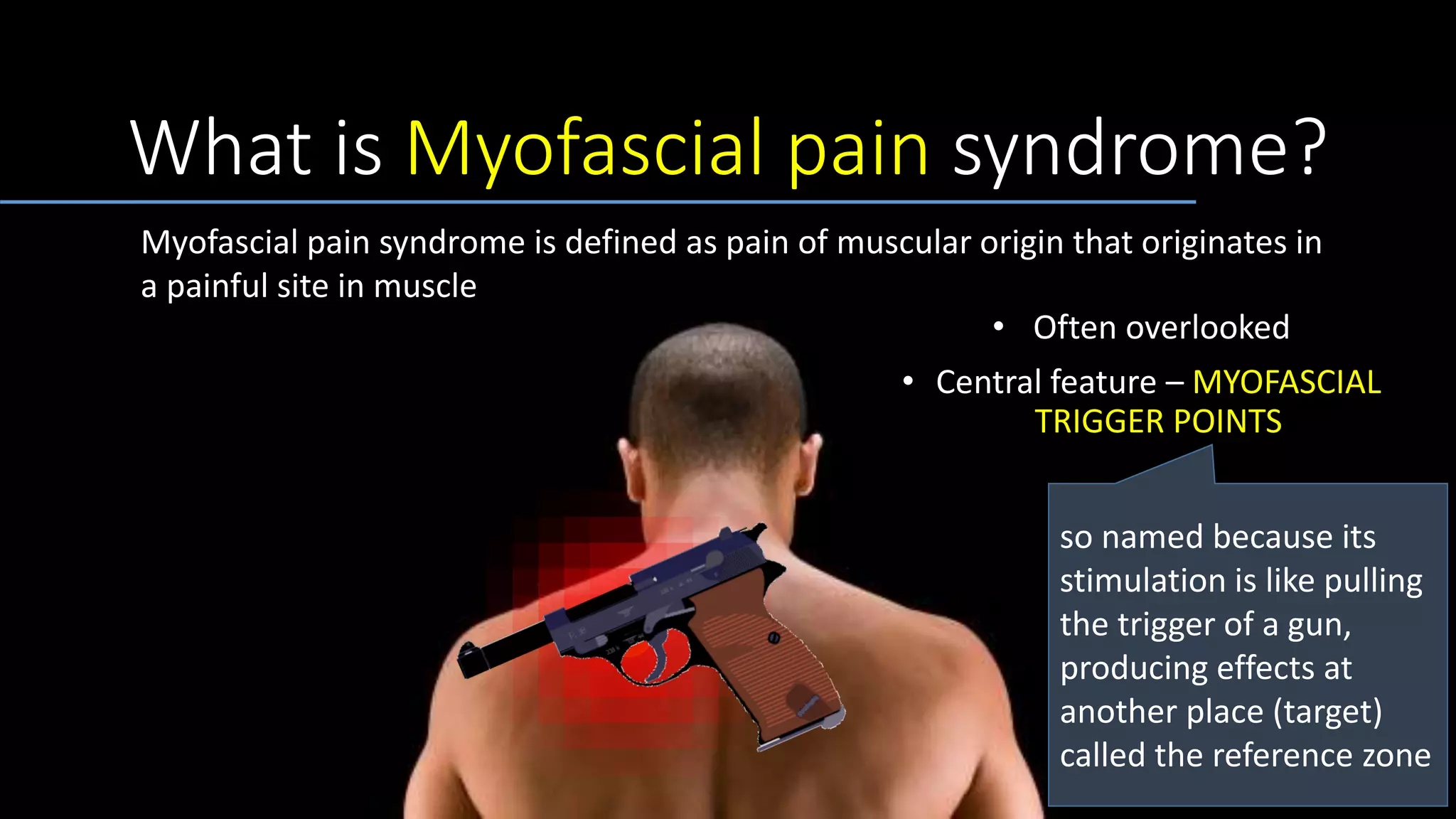
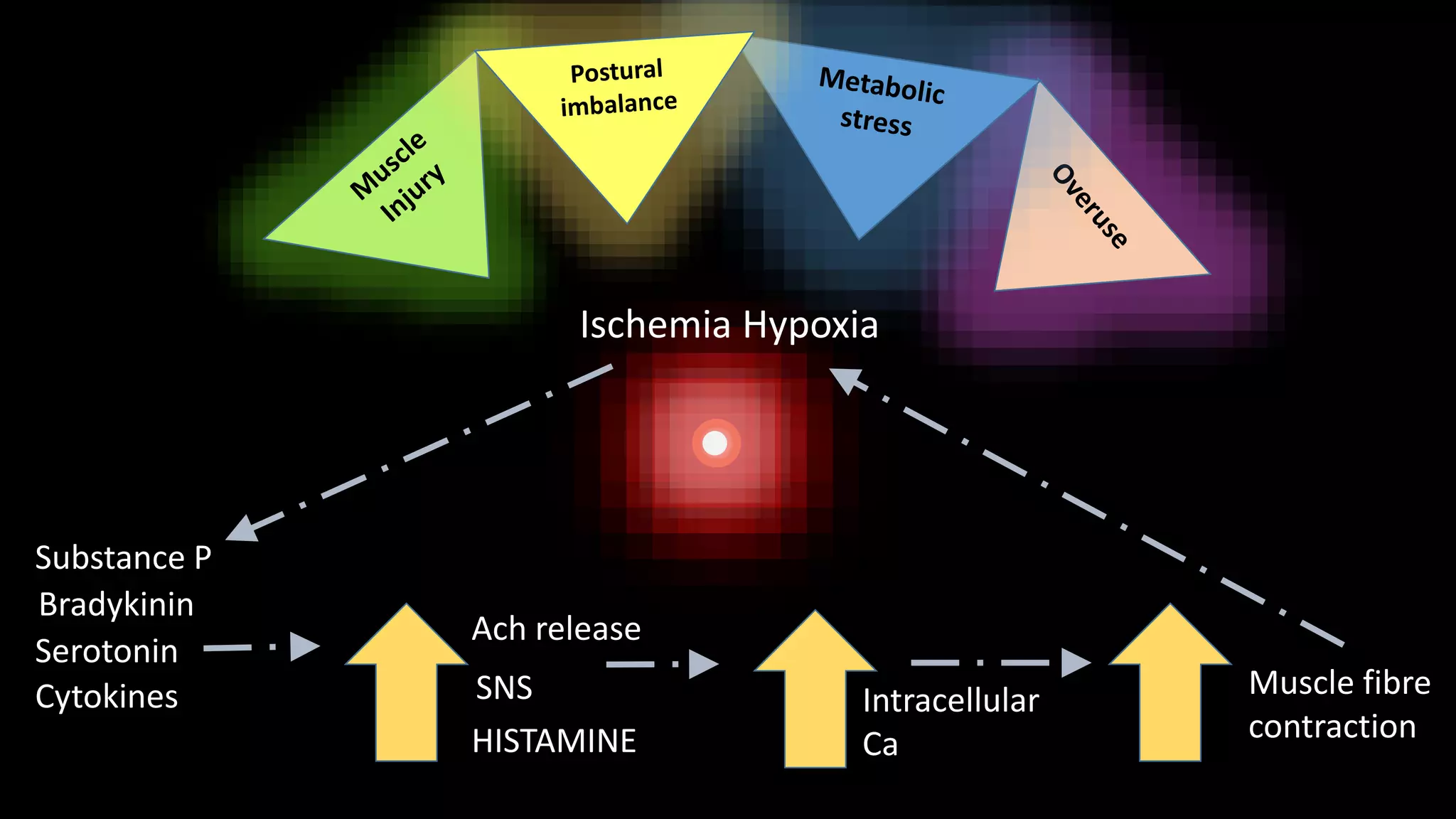
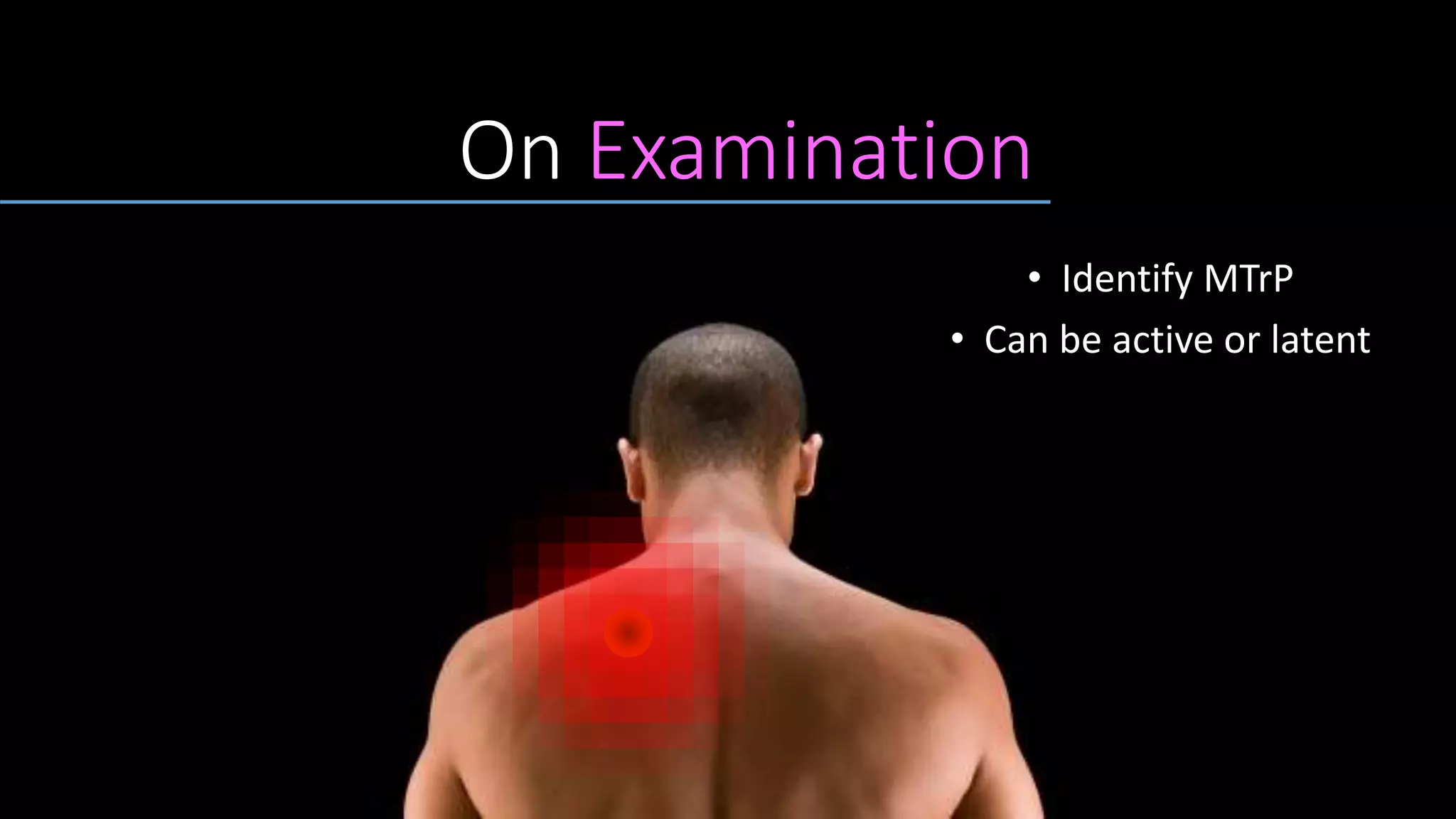
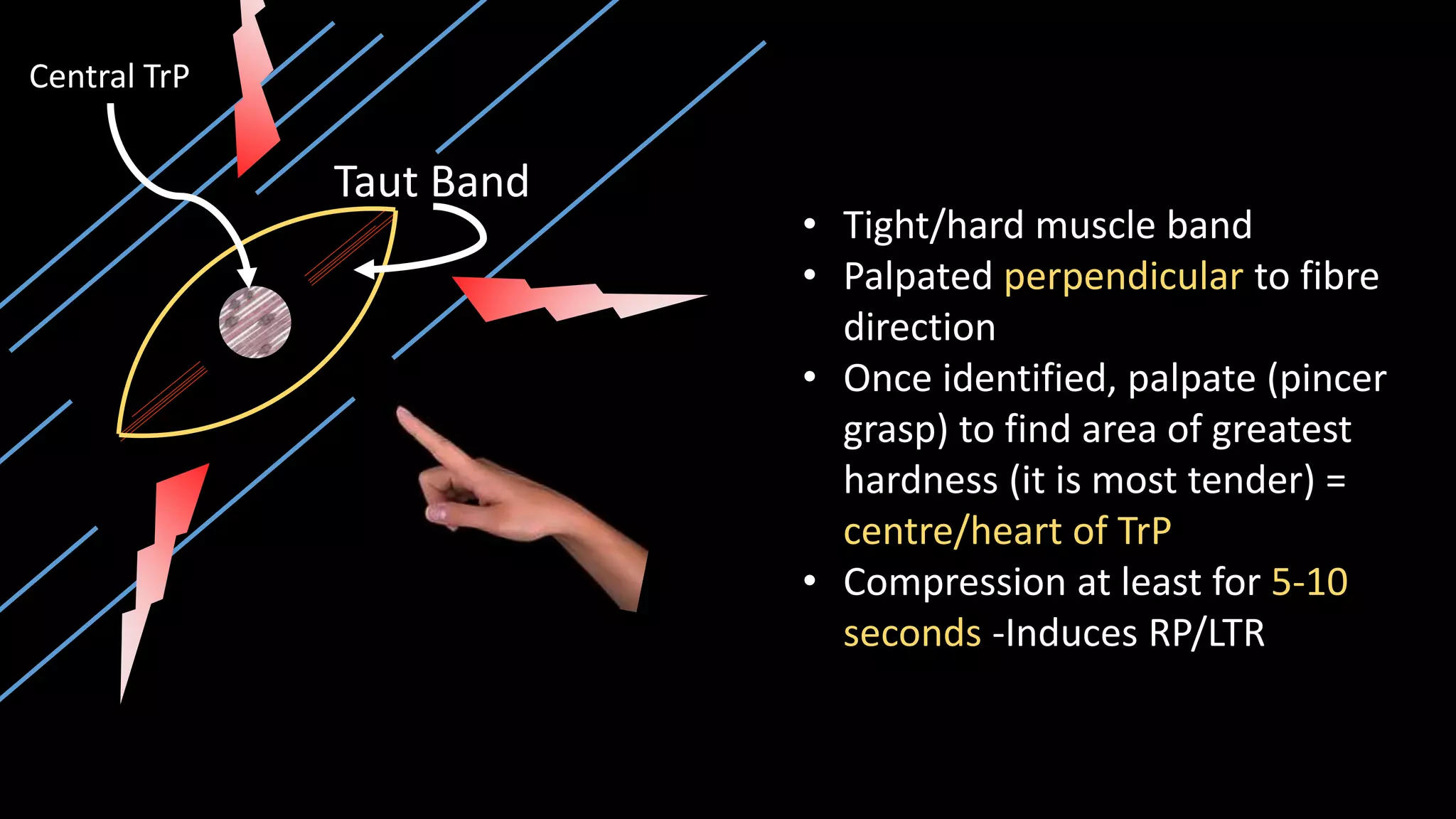
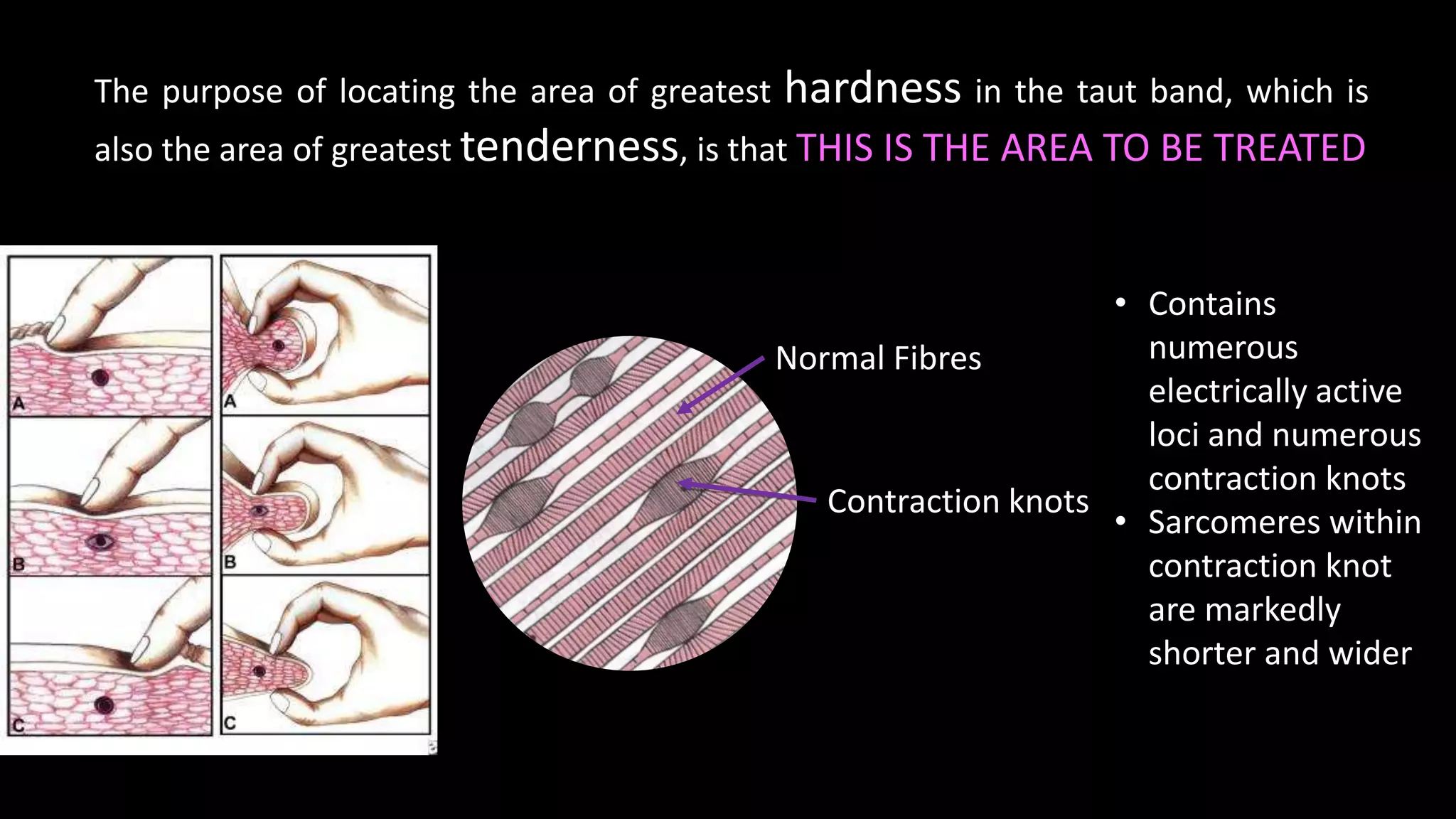
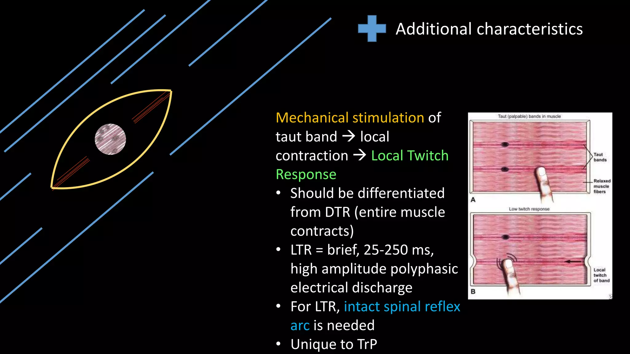
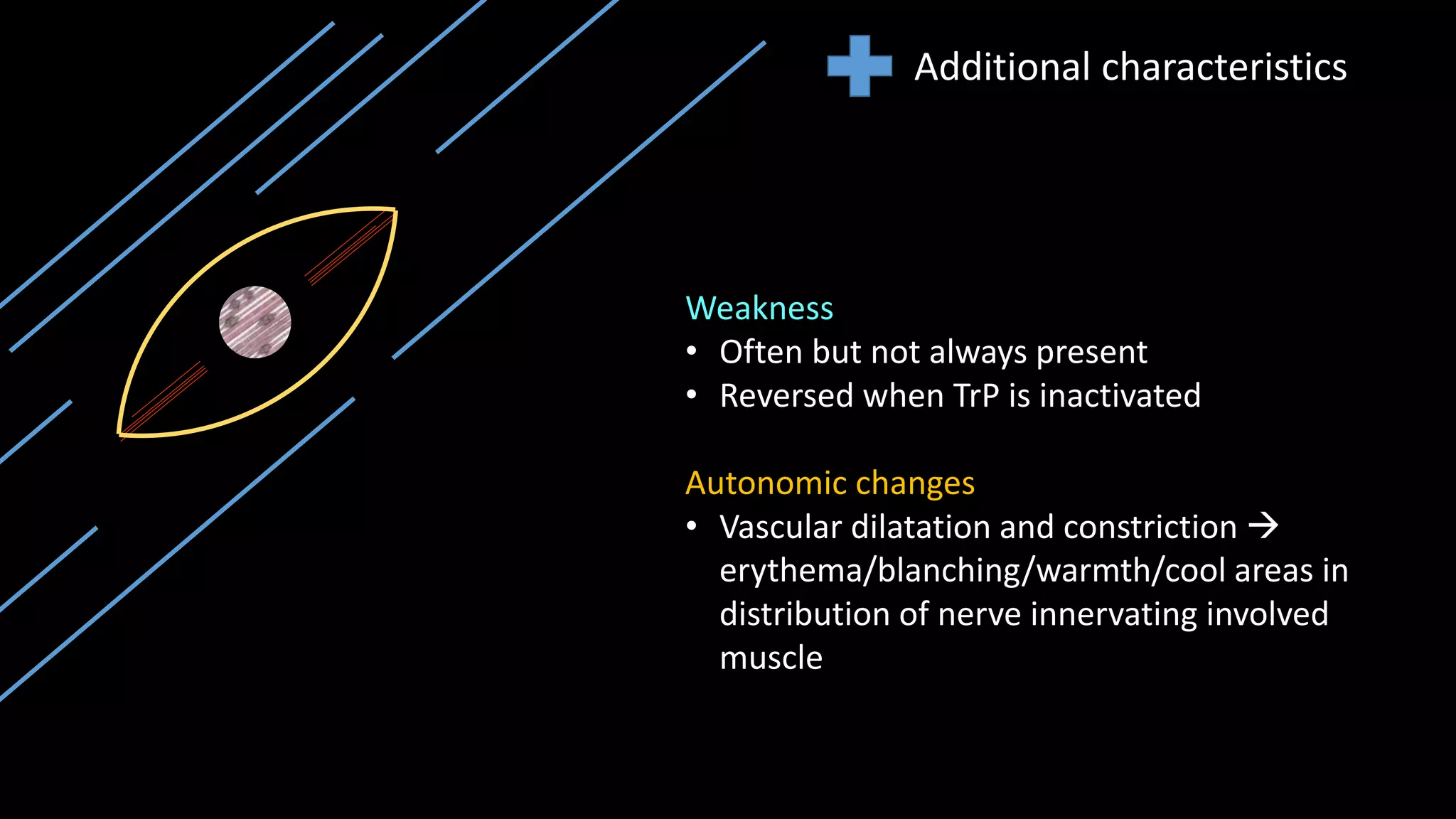
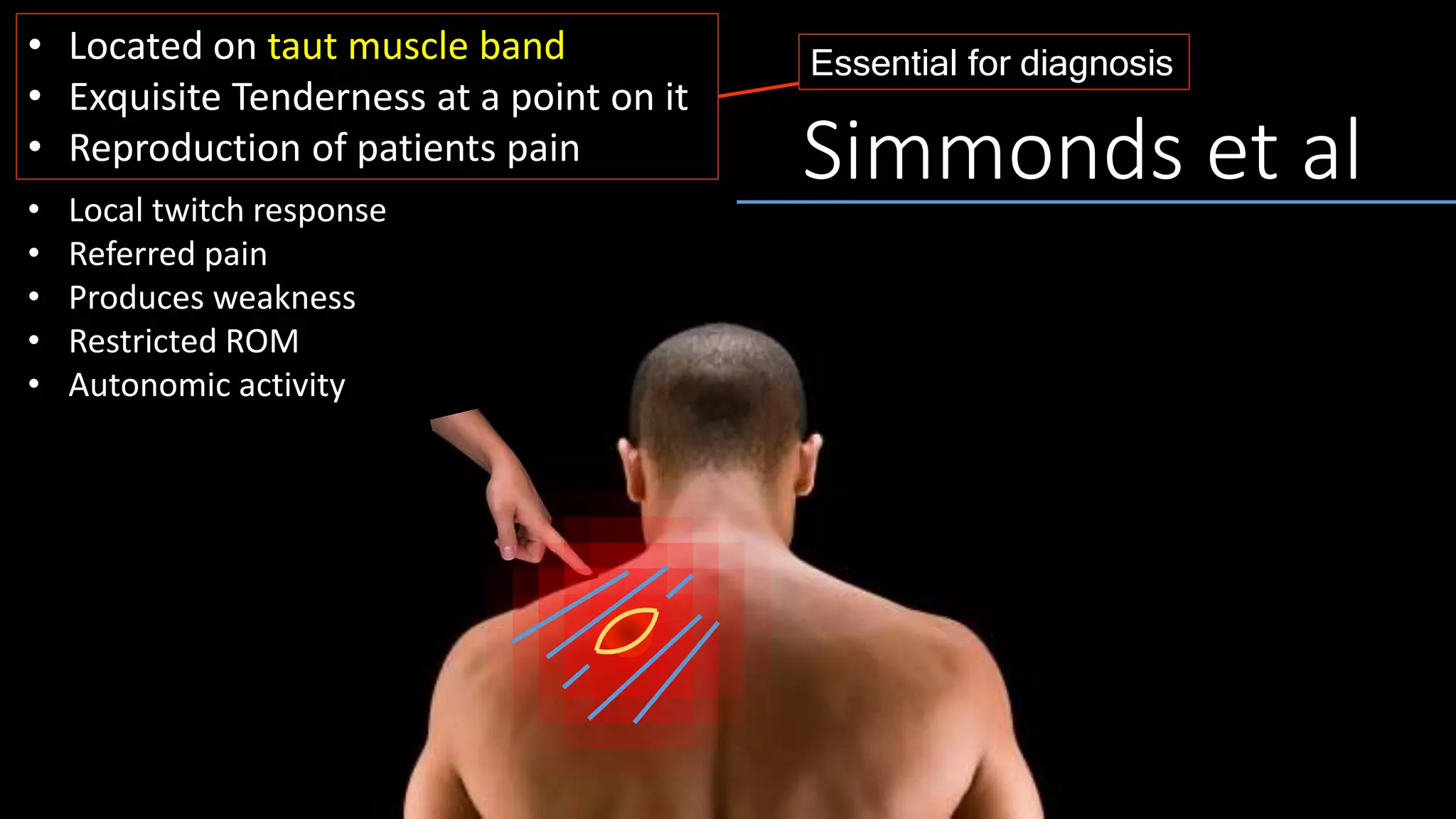
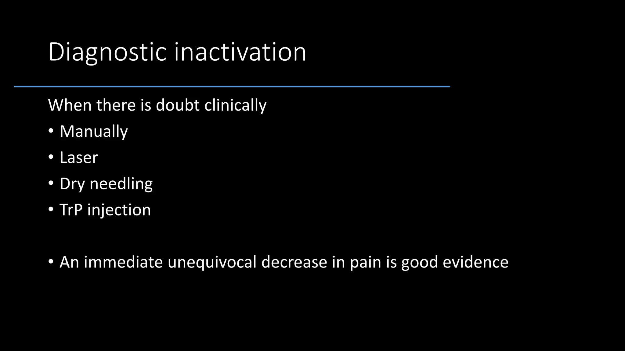
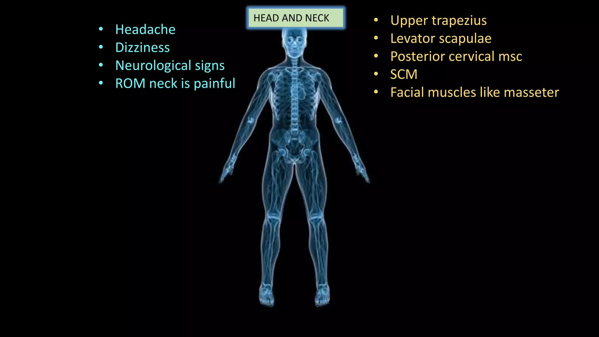
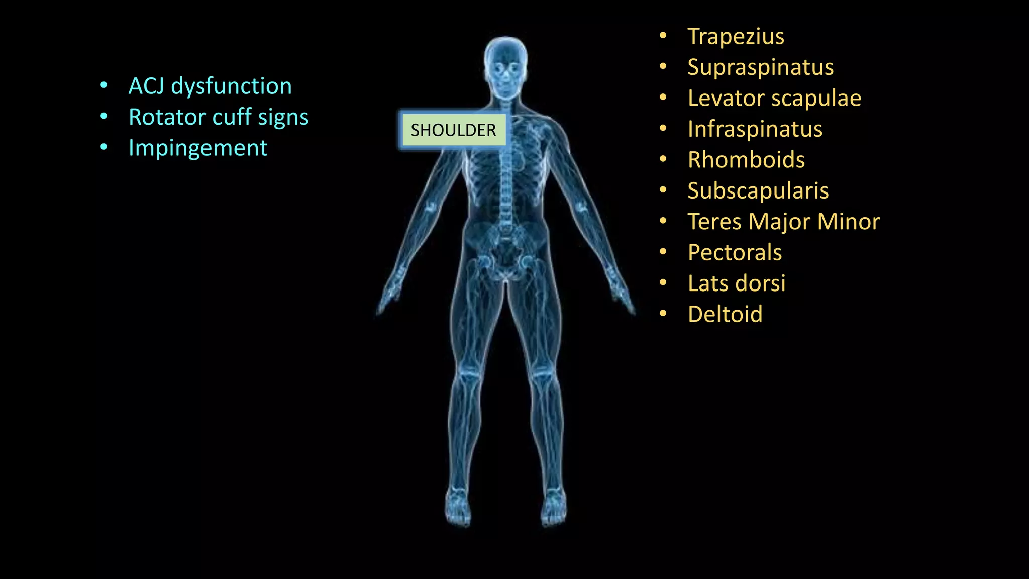
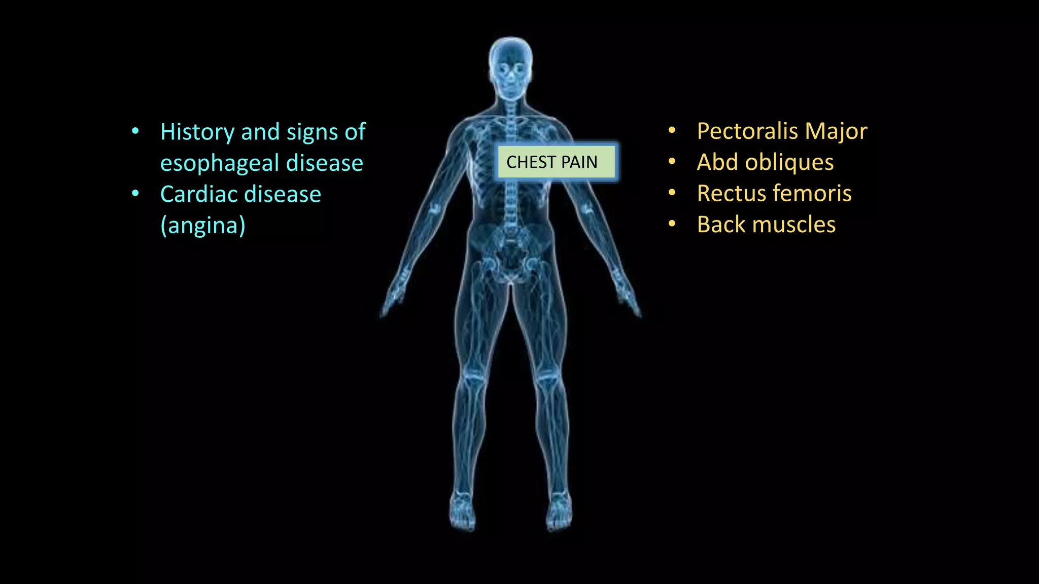
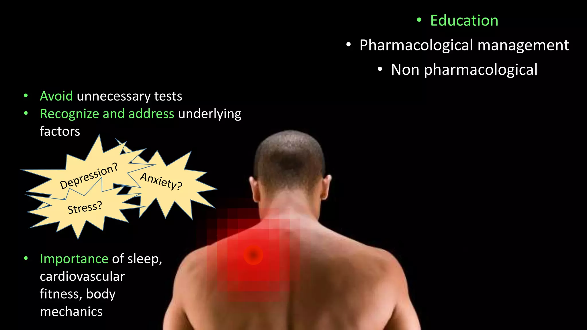
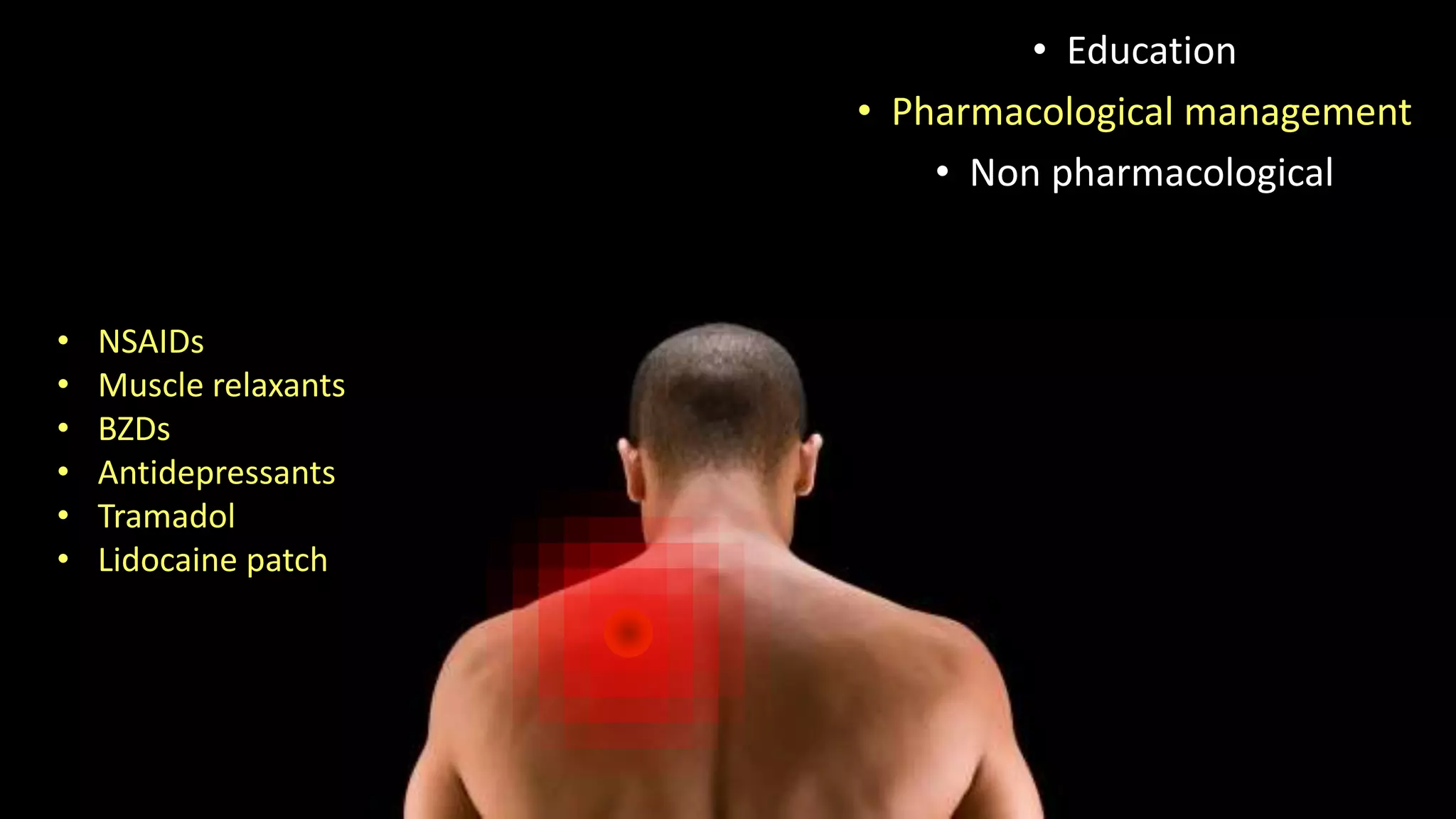
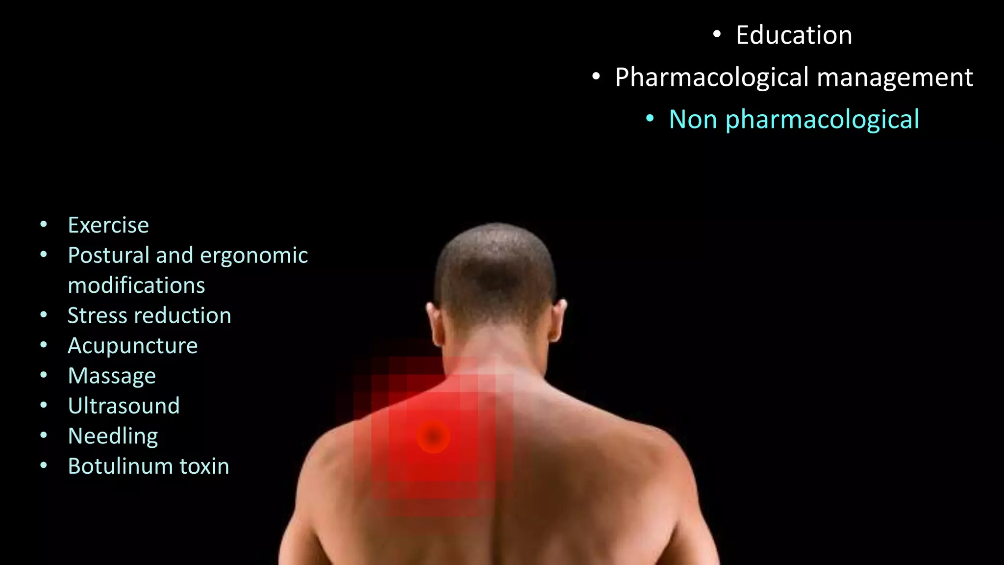
MYOFASCIAL PAIN
SYNDROME
Steffi Andrat
What is Myofascial pain syndrome?
• Often overlooked
• Central feature – MYOFASCIAL
TRIGGER POINTS
so named because its
stimulation is like pulling
the trigger of a gun,
producing effects at
another place (target)
called the reference zone
Myofascial pain syndrome is defined as pain of muscular origin that originates in
a painful site in muscle
History of Pain
• Acute/Chronic
• Dull, deep, aching
• Mimics radicular/visceral pain
• Often referred to head/ neck/
leg/ hip
Some predisposers
• History of remote injury
relevant
• Postural stress, muscle
imbalance, overuse
• Iron deficiency
• Hypothyroidism
• Low Vit D
• Low B12
• Parasitic infections
• Certain effects of sex hormones on pain
mechanisms
• Estradiol  modulates NMDA receptor in
dorsal horn  increasing nociceptive
response
• Estradiol  modulates excitability of
primary sensory afferent nerves
Gender
differences???
ETIOLOGY
How a trigger is formed
Mechanism for tenderness and referred pain
Substance P
Bradykinin
Serotonin
Cytokines
HISTAMINE
Intracellular
Ca
Muscle fibre
contraction
Ach release
SNS
Ischemia Hypoxia
Central sensitization
Central nervous system
modulates afferent
nociceptive activity
• SENSITIZATION to
peripheral noxious
stimuli in DORSAL
HORN NEURONS
Substance P  enhances
activation
Central sensitization
Dorsal horn neuron 
nociceptive impulses
rostrally 
activation of
somatosensory cortex 
interprets all input as
coming from receptive
field of that neuron
(expanded due to
sensitization)
On Examination
• Identify MTrP
• Can be active or latent
Taut Band
Central TrP
• Tight/hard muscle band
• Palpated perpendicular to fibre
direction
• Once identified, palpate (pincer
grasp) to find area of greatest
hardness (it is most tender) =
centre/heart of TrP
• Compression at least for 5-10
seconds -Induces RP/LTR
Normal Fibres
Contraction knots
The purpose of locating the area of greatest hardness in the taut band, which is
also the area of greatest tenderness, is that THIS IS THE AREA TO BE TREATED
• Contains
numerous
electrically active
loci and numerous
contraction knots
• Sarcomeres within
contraction knot
are markedly
shorter and wider
Additional characteristics
Mechanical stimulation of
taut band  local
contraction  Local Twitch
Response
• Should be differentiated
from DTR (entire muscle
contracts)
• LTR = brief, 25-250 ms,
high amplitude polyphasic
electrical discharge
• For LTR, intact spinal reflex
arc is needed
• Unique to TrP
Additional characteristics
Referred pain
Limited ROM
• Due to pain on lengthening affected muscle
• Examination gives clue about which muscle
has TrP
Additional characteristics
Weakness
• Often but not always present
• Reversed when TrP is inactivated
Autonomic changes
• Vascular dilatation and constriction 
erythema/blanching/warmth/cool areas in
distribution of nerve innervating involved
muscle
DIAGNOSIS
• Located on taut muscle band
• Exquisite Tenderness at a point on it
• Reproduction of patients pain
• Local twitch response
• Referred pain
• Produces weakness
• Restricted ROM
• Autonomic activity
Essential for diagnosis
Simmonds et al
Diagnostic inactivation
When there is doubt clinically
• Manually
• Laser
• Dry needling
• TrP injection
• An immediate unequivocal decrease in pain is good evidence
Objective identification
• MR elastography – differentiates tissues of varying densities
• Ultrasound – localizes hypoechoic elliptical focal areas
• EMG – Signature signal - persistant low amplitude, high frequency
discharge in the active TrP - spontaneous electrical activity
Lab investigations
• Not very usefu for diagnosis
• Can identify predisposers
• Anemia
• Hypothyroidism
• Vit D
• Vit B12
• Parasitic infections
Differential diagnosis of
REGIONAL PAIN SYNDROMES
HEAD AND NECK
• Headache
• Dizziness
• Neurological signs
• ROM neck is painful
• Upper trapezius
• Levator scapulae
• Posterior cervical msc
• SCM
• Facial muscles like masseter
SHOULDER
• ACJ dysfunction
• Rotator cuff signs
• Impingement
• Trapezius
• Supraspinatus
• Levator scapulae
• Infraspinatus
• Rhomboids
• Subscapularis
• Teres Major Minor
• Pectorals
• Lats dorsi
• Deltoid
CHEST PAIN
• History and signs of
esophageal disease
• Cardiac disease
(angina)
• Pectoralis Major
• Abd obliques
• Rectus femoris
• Back muscles
LOW BACK
• Spondyloarthropathis
• Spondylolisthesis
• PIVD
• Spinal stenosis
• Psoas
• Quadratus lumborum
• Paraspinals
• Abd obliques
• Rectii
PELVIS/HIP
• Internal organ disease
(painful bladder, IBS,
endometriosis)
• Radicular pain from LS spine
• Abdominal msc
• Psoas
• Quadratus lumborum
• Piriformis
• Adductors
• Hams (specially upper
Semitendinosis)
KNEE
• Intrinsic joint disease
• Radiculopathy
• Vastus medialis, lateralis
• Hamstring, gastrocnemius
ANKLE/FOOT
• Intrinsic joint disease
• Radiculopathy
• Anterior and posterior leg
muscles
• Gastroc-soleus
• Tibialis anterior
• Foot intrinsics
Treatment
• Education
• Pharmacological management
• Non pharmacological
• Avoid unnecessary tests
• Recognize and address underlying
factors
• Importance of sleep,
cardiovascular
fitness, body
mechanics
• NSAIDs
• Muscle relaxants
• BZDs
• Antidepressants
• Tramadol
• Lidocaine patch
• Education
• Pharmacological management
• Non pharmacological
• Exercise
• Postural and ergonomic
modifications
• Stress reduction
• Acupuncture
• Massage
• Ultrasound
• Needling
• Botulinum toxin
• Education
• Pharmacological management
• Non pharmacological
THANK YOU

Myofascial pain syndrome

  • 1.
  • 2.
    What is Myofascialpain syndrome? • Often overlooked • Central feature – MYOFASCIAL TRIGGER POINTS so named because its stimulation is like pulling the trigger of a gun, producing effects at another place (target) called the reference zone Myofascial pain syndrome is defined as pain of muscular origin that originates in a painful site in muscle
  • 3.
    History of Pain •Acute/Chronic • Dull, deep, aching • Mimics radicular/visceral pain • Often referred to head/ neck/ leg/ hip
  • 4.
    Some predisposers • Historyof remote injury relevant • Postural stress, muscle imbalance, overuse • Iron deficiency • Hypothyroidism • Low Vit D • Low B12 • Parasitic infections
  • 5.
    • Certain effectsof sex hormones on pain mechanisms • Estradiol  modulates NMDA receptor in dorsal horn  increasing nociceptive response • Estradiol  modulates excitability of primary sensory afferent nerves Gender differences???
  • 6.
    ETIOLOGY How a triggeris formed Mechanism for tenderness and referred pain
  • 7.
  • 8.
    Central sensitization Central nervoussystem modulates afferent nociceptive activity • SENSITIZATION to peripheral noxious stimuli in DORSAL HORN NEURONS Substance P  enhances activation
  • 9.
    Central sensitization Dorsal hornneuron  nociceptive impulses rostrally  activation of somatosensory cortex  interprets all input as coming from receptive field of that neuron (expanded due to sensitization)
  • 10.
    On Examination • IdentifyMTrP • Can be active or latent
  • 11.
    Taut Band Central TrP •Tight/hard muscle band • Palpated perpendicular to fibre direction • Once identified, palpate (pincer grasp) to find area of greatest hardness (it is most tender) = centre/heart of TrP • Compression at least for 5-10 seconds -Induces RP/LTR
  • 12.
    Normal Fibres Contraction knots Thepurpose of locating the area of greatest hardness in the taut band, which is also the area of greatest tenderness, is that THIS IS THE AREA TO BE TREATED • Contains numerous electrically active loci and numerous contraction knots • Sarcomeres within contraction knot are markedly shorter and wider
  • 13.
    Additional characteristics Mechanical stimulationof taut band  local contraction  Local Twitch Response • Should be differentiated from DTR (entire muscle contracts) • LTR = brief, 25-250 ms, high amplitude polyphasic electrical discharge • For LTR, intact spinal reflex arc is needed • Unique to TrP
  • 14.
    Additional characteristics Referred pain LimitedROM • Due to pain on lengthening affected muscle • Examination gives clue about which muscle has TrP
  • 15.
    Additional characteristics Weakness • Oftenbut not always present • Reversed when TrP is inactivated Autonomic changes • Vascular dilatation and constriction  erythema/blanching/warmth/cool areas in distribution of nerve innervating involved muscle
  • 16.
  • 17.
    • Located ontaut muscle band • Exquisite Tenderness at a point on it • Reproduction of patients pain • Local twitch response • Referred pain • Produces weakness • Restricted ROM • Autonomic activity Essential for diagnosis Simmonds et al
  • 18.
    Diagnostic inactivation When thereis doubt clinically • Manually • Laser • Dry needling • TrP injection • An immediate unequivocal decrease in pain is good evidence
  • 19.
    Objective identification • MRelastography – differentiates tissues of varying densities • Ultrasound – localizes hypoechoic elliptical focal areas • EMG – Signature signal - persistant low amplitude, high frequency discharge in the active TrP - spontaneous electrical activity
  • 20.
    Lab investigations • Notvery usefu for diagnosis • Can identify predisposers • Anemia • Hypothyroidism • Vit D • Vit B12 • Parasitic infections
  • 21.
  • 22.
    HEAD AND NECK •Headache • Dizziness • Neurological signs • ROM neck is painful • Upper trapezius • Levator scapulae • Posterior cervical msc • SCM • Facial muscles like masseter
  • 23.
    SHOULDER • ACJ dysfunction •Rotator cuff signs • Impingement • Trapezius • Supraspinatus • Levator scapulae • Infraspinatus • Rhomboids • Subscapularis • Teres Major Minor • Pectorals • Lats dorsi • Deltoid
  • 24.
    CHEST PAIN • Historyand signs of esophageal disease • Cardiac disease (angina) • Pectoralis Major • Abd obliques • Rectus femoris • Back muscles
  • 25.
    LOW BACK • Spondyloarthropathis •Spondylolisthesis • PIVD • Spinal stenosis • Psoas • Quadratus lumborum • Paraspinals • Abd obliques • Rectii
  • 26.
    PELVIS/HIP • Internal organdisease (painful bladder, IBS, endometriosis) • Radicular pain from LS spine • Abdominal msc • Psoas • Quadratus lumborum • Piriformis • Adductors • Hams (specially upper Semitendinosis)
  • 27.
    KNEE • Intrinsic jointdisease • Radiculopathy • Vastus medialis, lateralis • Hamstring, gastrocnemius
  • 28.
    ANKLE/FOOT • Intrinsic jointdisease • Radiculopathy • Anterior and posterior leg muscles • Gastroc-soleus • Tibialis anterior • Foot intrinsics
  • 29.
  • 30.
    • Education • Pharmacologicalmanagement • Non pharmacological • Avoid unnecessary tests • Recognize and address underlying factors • Importance of sleep, cardiovascular fitness, body mechanics
  • 31.
    • NSAIDs • Musclerelaxants • BZDs • Antidepressants • Tramadol • Lidocaine patch • Education • Pharmacological management • Non pharmacological
  • 32.
    • Exercise • Posturaland ergonomic modifications • Stress reduction • Acupuncture • Massage • Ultrasound • Needling • Botulinum toxin • Education • Pharmacological management • Non pharmacological
  • 33.