This document discusses strategies for improving pedicle screw fixation in osteoporotic bone. It describes the advantages of pedicle screw constructs but notes their risk of failure in osteoporosis due to poor bone quality compromising screw fixation. It then presents a novel technique of using fenestrated, cement-augmented pedicle screws that allows cement injection through the screw after placement to strengthen fixation. Comparative studies show this technique increases pullout strength while decreasing the risks of cement leakage compared to traditional cement augmentation. The document concludes by discussing other approaches like expansive screws, bicortical purchase, and multiple levels of fixation that can further improve construct stability in osteoporotic patients.
Introduction to spinal arthrodesis using pedicle screws in osteoporosis presented by Mohamed Mohi Eldin, MD.
Discusses the advantages of pedicle screw constructs: three-column fixation, short-segments instrumentation, and maintaining sagittal alignment.
Emphasizes that the key to performance is the strength of attachment to the spine, highlighting the need for solid fixation in quality trabecular bone.
Describes how aging affects bone mineral density (BMD), a key factor in spinal instrumentation failure, noting that poor BMD compromises screw fixation.
Stresses the inevitability of needing spinal decompression and stabilization in cases of osteoporosis and poor bone quality.
Introduction to the topic of secondary osteoporosis and its relevance.
Performance issues in osteoporotic patients, including complications like screw loosening and high failure risk due to screw pullout.
Methods to improve screw fixation in osteoporotic bone: cement augmentation, multiaxial screws, bicortical and iliac screws, among others.
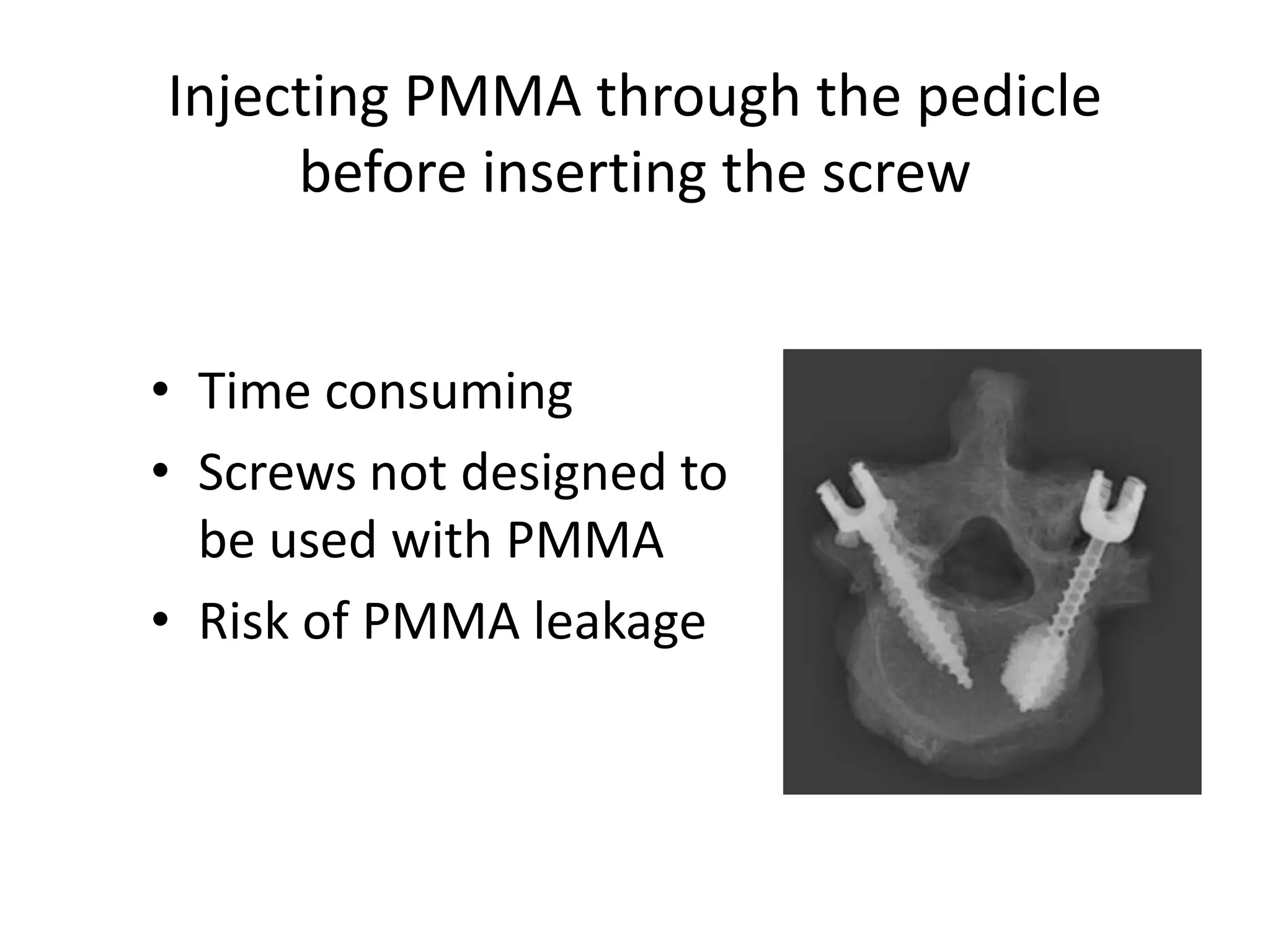
Introduction of canulated, fenestrated, augmented screws and old attempts to enhance fixation through cement diffusion before screw insertion.
Describes complications of injecting PMMA, including thermal damage, difficulty in removal, and neurologic risks.
Proposes a novel concept of injecting PMMA through fenestrated screws to optimize cement distribution.
Discusses surgical planning for different techniques and shares important positions to avoid leakage during cement injection.
Details techniques for cement delivery, including viscosity management and fluoroscopic control to prevent leakage.
Combining techniques: discusses cement volumes for thoracic and lumbar screws with focus on safe injection practices.
Underlines importance of screw alignment and positioning for effective cement distribution and avoiding breaches.
Conducts comparative analysis between augmented fenestrated screws and standard screws, highlighting strength and safety.
Evaluates clinical safety comparing fenestrated vs. traditional techniques, emphasizing reduced risk of complications.
Analyzes cement leakage in traditional techniques vs. operatively efficient fenestrated technique, which allows easier screw placement.
Highlights the benefits of the fenestrated technique in terms of efficiency and presents tips to prevent extravasation.
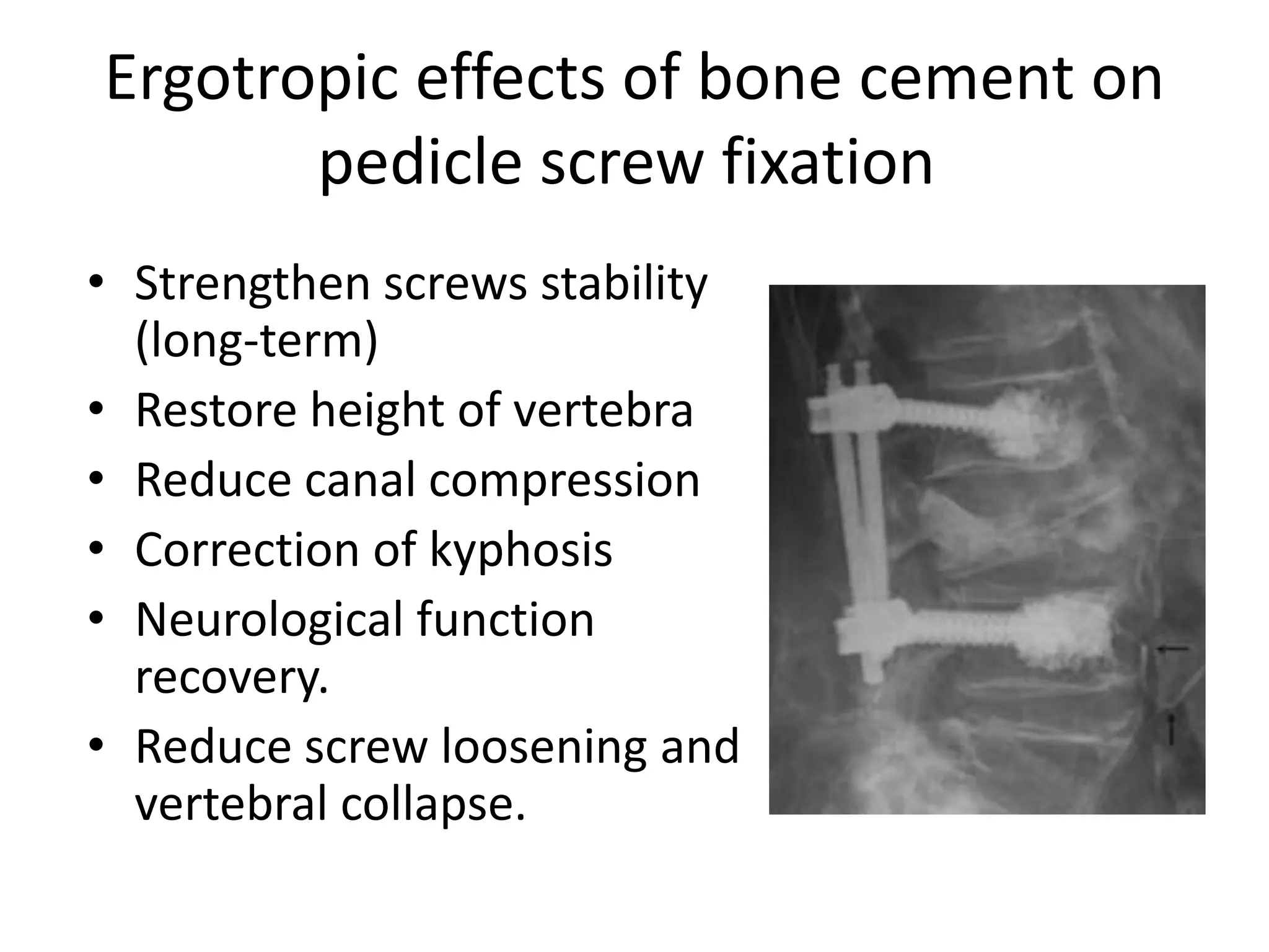
Discusses the ergotropic effects of bone cement, including stability strengthening and other positive outcomes as compared to extended instrumentation.
Presents how expansive pedicle screws function and stresses the role of cement augmentation in improving pullout strength.
Explains bicortical screw benefits for improving strength and the associated risks of anterior structure damage.
Focuses on hydroxyapatite coating on screws to enhance screw-bone contact and pullout strength.
Explores the potential of using double screws in cadaver studies to improve fixation in osteoporotic vertebrae.
Discusses the benefit of multilevel constructs and extension of fusion to reduce failure risks.
Presents various case studies including spondylolisthesis and burst fractures, showcasing surgical techniques used.
Spinal Arthrodesis Using
PedicleScrews in
Osteoporosis
Mohamed Mohi Eldin, MD,
Professor of Neurosurgery,
Faculty of Medicine,
Cairo University,
Egypt.
Performance of PedicleScrew Systems
The key is strength of attachment to spine.
Solid fixation (when screws are placed in dense,
good-quality trabecular bone)
4.
Aging population
• Bonemineral density (BMD) is one of main
factors related to spinal instrumentation
failure.
• In vitro: poor BMD compromises screw
fixation.
5.
For a spinesurgeon
It is unavoidable to require
spinal decompression &
stabilization for osteoporosis
or other decreased bone
quality
Performance of pediclescrews
In osteoporotic patients
potential complications
screw loosening,
migration, or pullout,
compromising outcome
Screw-bone interface
stripped easily during
screw tapping and
insertion
8.
Pedicle screws useis relatively
contraindicated in osteoporosis
because of:
• High risk of failure (screw pullout)
• Damage caused by such pullouts can
complicate revision surgeries.
9.
How to improvepedicle screw fixation
in osteoporotic bone
1. Augmentation with cement
2. Multiaxial expandable pedicle screw
3. Bicortical sacral screws for the sacrum
4. Bilateral iliac screws for the pelvis
5. Hydroxyapatite Coating of screws
6. Double screws (Only in cadavers)
7. Multiple levels
8. Increase points of fixation
Old Attempts
To enhancepedicle screw fixation
Passive diffusion of cement
into the vertebral body and
pedicle
immediately prior to screw
insertion
12.
Injecting PMMA throughthe pedicle
before inserting the screw
• Time consuming
• Screws not designed to
be used with PMMA
• Risk of PMMA leakage
13.
PMMA traditional techniqueRisks
• Extravasation (fractured
cortex)
• Thermal damage,
• Difficult removal,
• Potential for major
neurologic compromise
• Rare fatal fat embolism
14.
However
• Time couldbe diminished
• Avoidance of extrusion could be done by
Altering delivery of PMMA
through a fenestrated pedicle screw.
15.
Cannulated Screw withDistal Fenestrations
(a novel-concept)
After screw insertion, cement can be injected
and will distribute evenly around
the thread of the screw
16.
The cement isextruded
through the fenestrations
to fill the spaces inside the
osteoporotic cancellous
bone;
Under fluoroscopic guidance
17.
Depending on thesurgical plan
Either
– A pure bilateral percutaneous
pedicle screw arthrodesis
– A combination of unilateral
percutaneous with a
contralateral miniopen
(modified Wiltse).
18.
For the PurePercutaneous
Fenestrated Screw Placement
The position of holes,
as far as possible from
posterior wall
(to prevent leakage)
19.
After augmentation, changeof screw
position is no longer possible
Trial of placement of the
rod should be done
before cement
injection to avoid
positioning issues after
cement injection.
20.
• PMMA bonecement is
delivered through
– the cement cannula within
the screws
– Cement delivery system
connected by connector.
• To prevent cement
leakage, the injection is
done in a higher viscosity
state (started 5 minutes
after mixing).
21.
• Injection mustbe done
under fluoroscopic
control to immediately
stop the injection in
case of cement
extravasations
– anterior,
– posterior, or
– into an adjacent disc
For combination ofunilateral percutaneous
with a contralateral Miniopen Techniques
When a central canal
decompression or
an interbody fusion
(LIF) is planned
24.
Cement Volume
injected intoeach screw
• Varies from 1.5 to 3mL.
– The ideal amount is 2mL.
– The safer amount
• 1.0 cc for the thoracic spine
• 1.5 cc for the lumbar spine
• Greater volume did not
lead to higher pullout
strength.
25.
An optimal positioningof the
fenestrated pedicle screws
• The length of screw is
important, as far as possible
from the posterior wall.
• Placed in middle of pedicle to
avoid cortical breaches
• An optimal alignment with
pedicle with good convergent
trajectory.
26.
The Location &Dispersion
of Cement
• The fenestrations,
whether partial or
full, led to a more
optimal distribution
of cement inside the
vertebrae
27.
Typical bone cement
distributionin lumbar
spine
Micro-CT showing
the interdigitation of
the bone cement
within the vertebral
body
28.
When compared withstandard pedicle
screws without PMMA
Augmented fenestrated
screws showed increased
pullout strength in
osteoporosis
29.
When Compared to
TraditionalTechnique
• Cement injection
increases pull-out
strength (200%) in both
• The fenestrated cement
technique required
more power until failure
Cross-section
30.
Comparing Clinical Safety
•The fenestrated technique
decrease the risk of nerve
injury significantly
– small numbers of pedicle wall
violations
– no extrusion of cement into
spinal canal
• Medial wall violations in
traditional delivery technique
with PMMA extrusion into
the canal.
31.
Comparing location ofcement
• In the fenestrated technique,
the cement confined to
vertebral body with NO
proximal migration (screw as
a plug)
• Even in the presence of
pedicle wall violation during
insertion.
32.
Cement Leakage
in traditionaldelivery technique
The PMMA is pushed against the walls of the pedicle
during screw insertion, allowing cement to extrude
from any violation into spinal canal.
33.
Operative efficiency ofthe fenestrated
delivery technique
• In traditional technique,
– Number of pedicle screws is
limited by how fast the cement
hardens vs. how fast a surgeon
can place screws by hand.
• In fenestrated technique,
– all screws are placed prior to
mixing the cement so cement is
injected through pre-placed
screws much easier.
Avoid breakage ofthe cement bridges
between the screw & bone
• Before injecting cement:
– Fixation system locked
– Rods tested in position
• After injecting cement:
– No torsion movement
should be applied to
screws
38.
Ergotropic effects ofbone cement on
pedicle screw fixation
• Strengthen screws stability
(long-term)
• Restore height of vertebra
• Reduce canal compression
• Correction of kyphosis
• Neurological function
recovery.
• Reduce screw loosening and
vertebral collapse.
39.
Cement augmentation versus
extendedinstrumentation
Lengthening
instrumentation
results in
higher increase of stability
during fatigue testing
in osteoporotic spine
compared with cement
augmentation.
Expansive screws afterexpansion
(A) 6-slit screw with
16-mm EEL
(B) 6-slit screw with
22-mm EEL.
44.
• Increase inboth the number of slits and the
extent of screw expansion had little impact on
the screw-anchoring strength.
• Cement augmentation is the most influential
factor for improving screw pullout strength for
expansive pedicle screws.
The Effects of Screw Design and
Cement Augmentation
45.
Various cemented screws
afterpullout tests
Left to right:
• solid,
• 4-slit with 16-mm,
• 4-slit with 22-mm,
• 6-slit with 16-mm,
• 6-slit with 22-mm.
46.
Bicortical purchase
• Thecortex is stronger than cancellous bone
in normal and osteoporotic vertebrae,
• Logically stronger than cortical-cancellous
purchase,
• Improve pullout strength from 20% to 50%,
depending on
– patient’s vertebrae,
– screw dimensions,
– screw type
47.
Bicortical screw increasesrisk of
damage to various anterior structures
usually used only at the S1 level (avoiding
damage to the aorta and vena cava)
48.
Hydroxyapatite Coating ofscrews
• Improving pedicle screw-bone contact,
• Improving bone ingrowth, and mineralization
• Increasing screw pullout strength
• Require significantly greater insertional torque
than uncoated screws
49.
Double screws
(Only incadaveric studies)
Because pedicles of osteoporotic vertebrae are
often larger than those of non-osteoporotic
vertebra, it may be possible to implant two
small-diameter pedicle screws into one
pedicle to improve fixation.
50.
Multiple levels andpoints of fixation
• Cement augmentation of
most rostral pedicle screws
can also help avoid failure
in multilevel constructs
• Extension of the fusion to
the sacrum/ilium can
reduce the potential high
risk for failure of constructs
ending with pedicle screws
at L5.