Downloaded 103 times


This document summarizes a seminar on peptic ulcer disease. It defines peptic ulcers, classifies them as acute or chronic, and discusses their etiology, including H. pylori infection and stress factors. It covers the pathogenesis of ulcers, clinical features, diagnosis including tests for H. pylori, and treatment using proton pump inhibitors, H2 receptor antagonists, and antibiotics. It also discusses complications, factors affecting treatment success, adverse drug reactions, drug interactions, and patient counseling.
Seminar introduction on Peptic Ulcer Disease led by Walid S Momin from Pharmacy Practice.
Peptic ulcers are mucosal degeneration areas in the gastrointestinal tract exposed to acid.
Peptic ulcers classified into acute (multiple small erosions) and chronic (gastric or duodenal).
Peptic ulcer etiology includes stress, H-Pylori infection, medications, and various irritants.
H-Pylori infection can lead to gastritis and ultimately peptic ulcers.
Details on acid secretion mechanisms and factors in the pathogenesis of peptic ulcers.
Pain occurring 1-3 hours after meals, anorexia, nocturnal pain, and potential nausea.
Diagnosis of H-Pylori through non-invasive tests, invasive techniques, and biopsy methods.
Techniques for visualizing erosions and bleeding sites, including barium contrast studies.
Non-pharmacological approaches and pharmacological therapy classification for ulcers.Risks of bleeding ulcers and late complications requiring treatments like somatostatin.
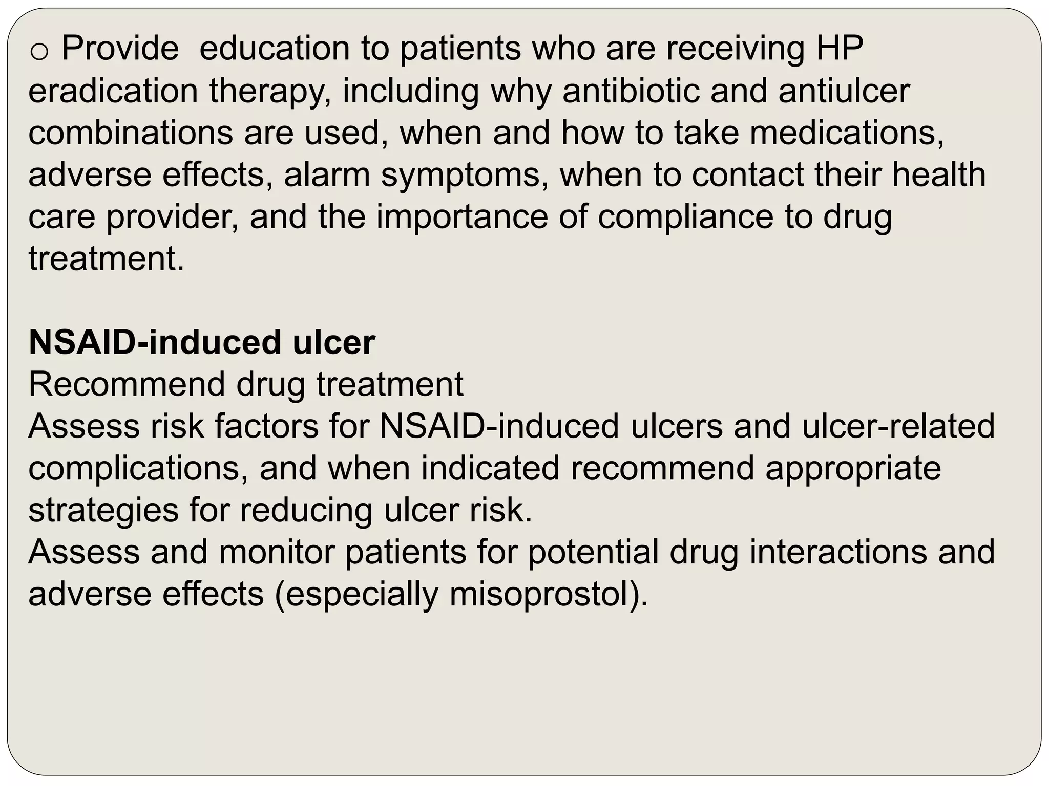
Guidelines for monitoring H-Pylori treatment, patient education, and prevention strategies.
Factors contributing to unsuccessful eradication include patient compliance and antibiotic resistance.
Side effects of various treatments including PPIs, H2 antagonists, and their clinical implications.
Key references for further reading on pharmacotherapy and clinical management of ulcers.


