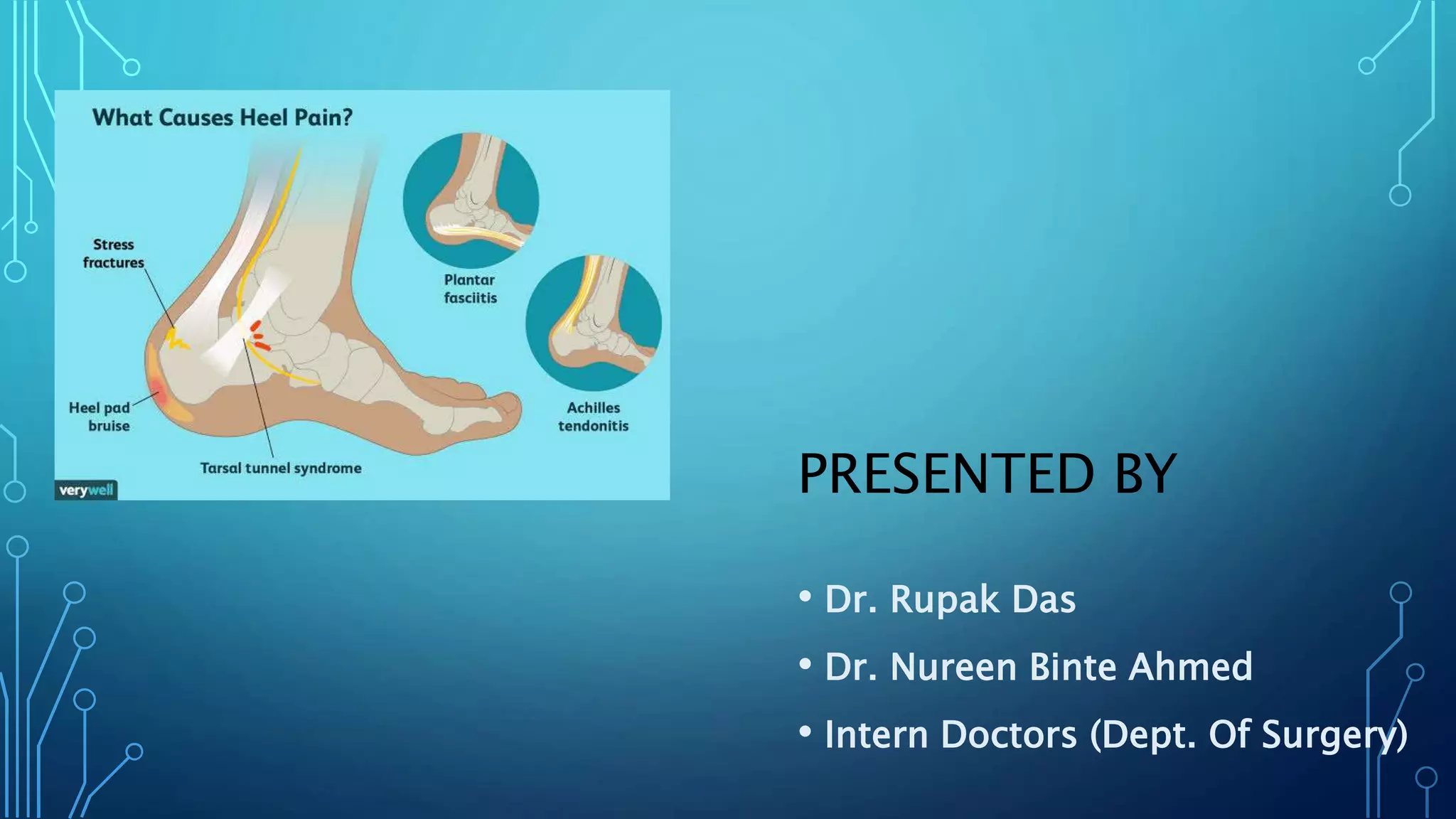
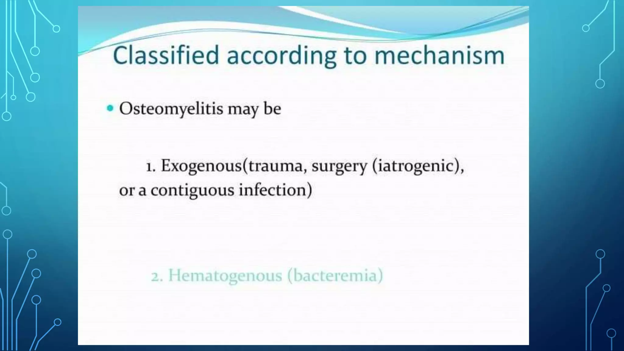
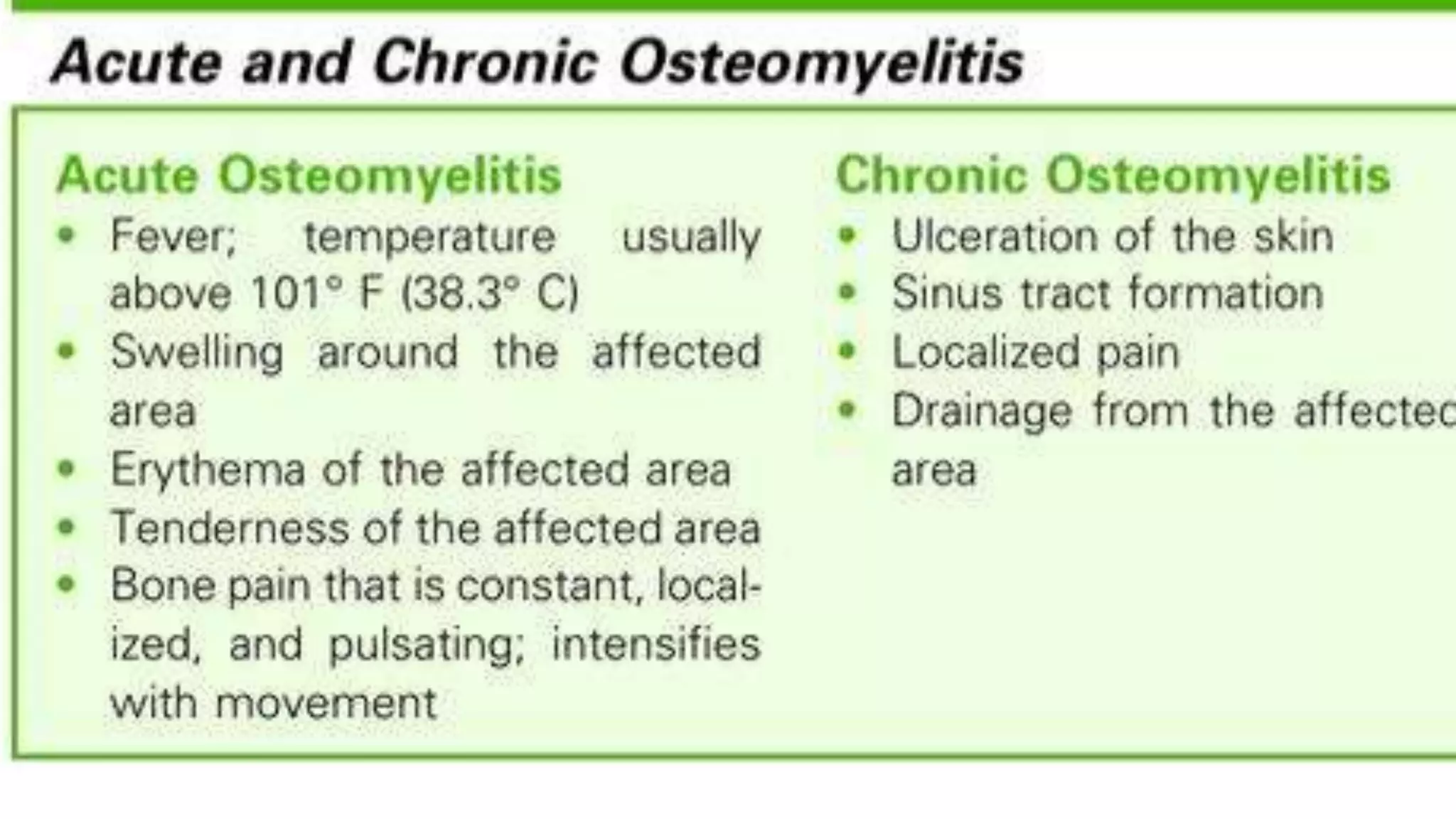
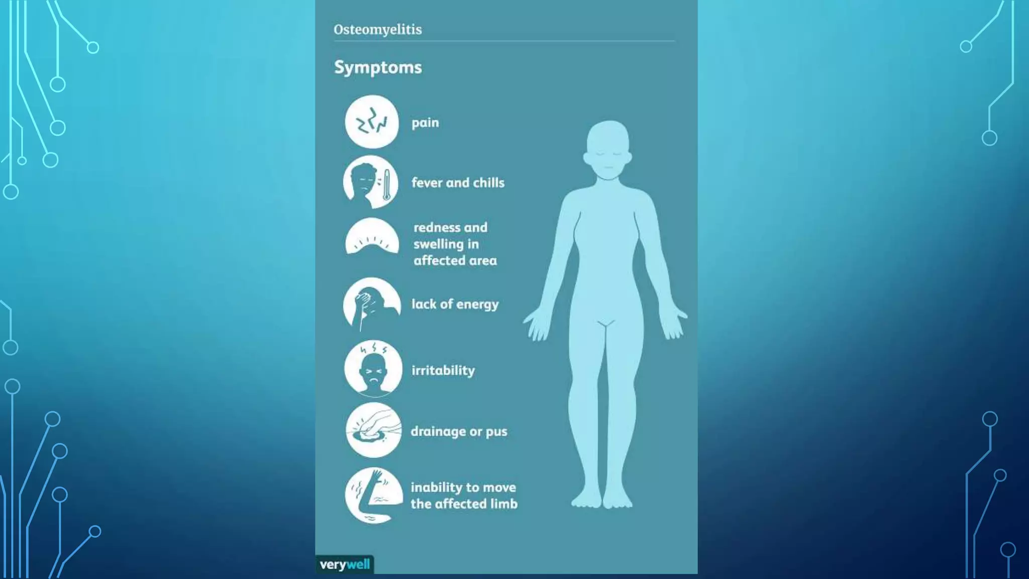
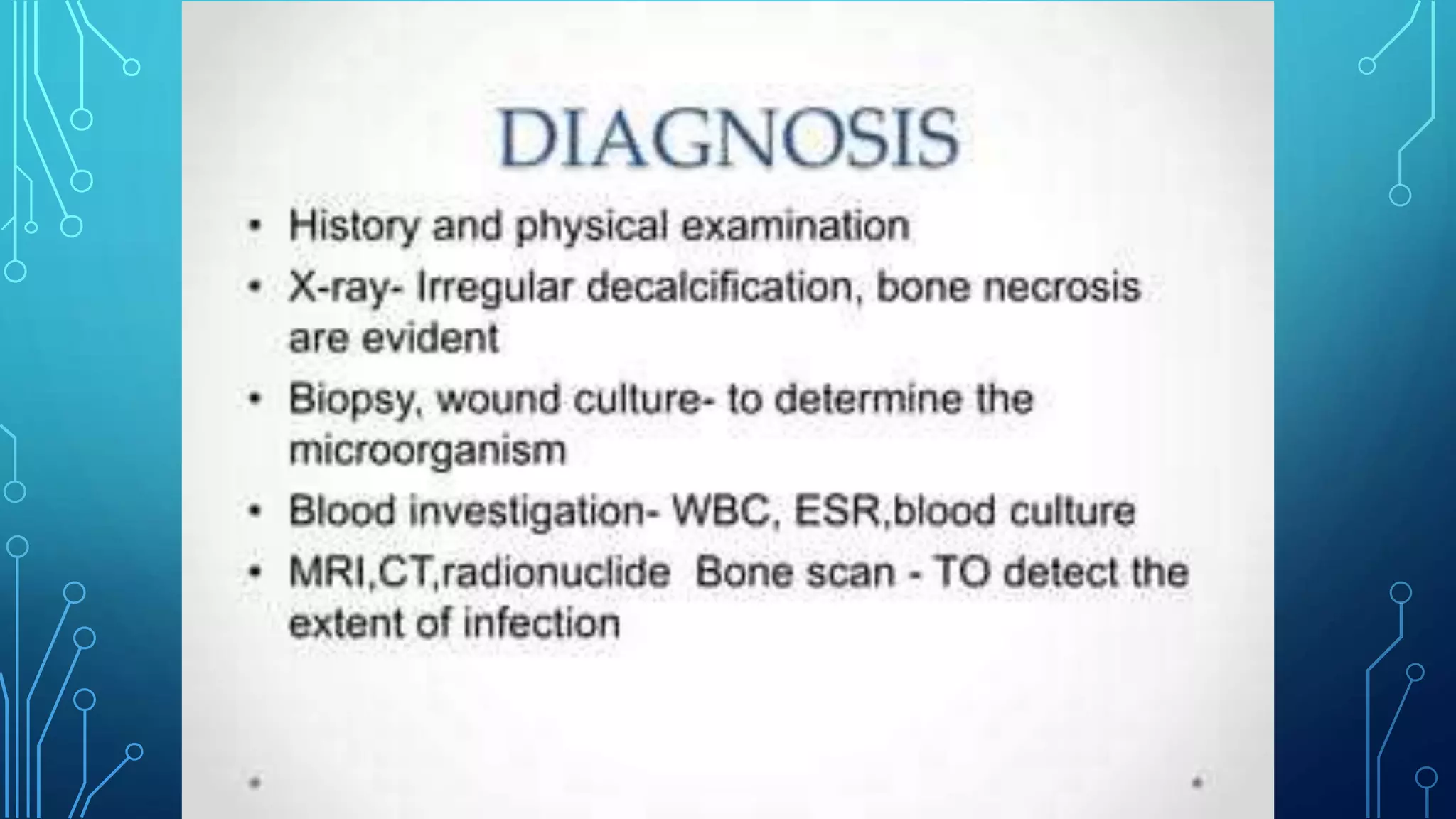
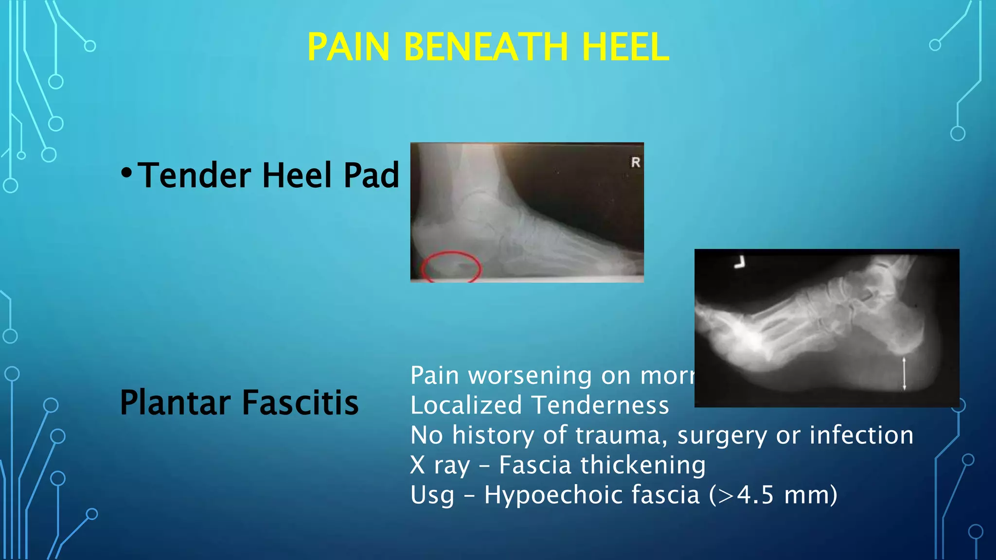
WELCOME
TODAY’S TOPIC
PAINFUL HEEL
PRESENTED BY
• Dr. Rupak Das
• Dr. Nureen Binte Ahmed
• Intern Doctors (Dept. Of Surgery)
PAINFUL HEEL
The causes of painful heel are conveniently classified according to the site of the pain .
Pain within the heel
Disease of the calcaneus (osteomyelitis, tumour, Paget's disease.)
Arthritis of the subtalar joint
Pain beneath the heel
Tender heel pad, Plantar fasciitis
Pain behind the heel
Rupture of the calcaneal tendon, Calcaneal paratendinitis, Post-calcaneal bursitis
,Calcaneal apophysitis
PLANTAR FASCITIS
• This is an annoying and/painful condition that limits function.
There is pain and tenderness in the sole of the foot, mostly
under the heel, with standing or walking.
• condition usually comes on gradually, without any clear
incident or injury, but sometimes there is a history of sudden
increase in sporting activity, or a change of footwear, sports
shoes or running surface. There may be an associated tightness
of the Achilles tendon. The pain is often worse when first
getting up in the morning.
PATHOLOGY
• The plantar fascia or aponeurosis is a dense fibrous structure
that originates from the calcaneum, deep to the heel fat pad,
and runs distally to the ball of the foot, with slips to each toe.
The plantar fascia stiff- ens and becomes less pliable with age.
The fascia is probably not actually inflamed in this condition, at
least not beyond the first week or two of onset. There may be
micro-tears in the fascia, and the fascia thick- ens. The
condition is sometimes associated with inflam- matory
disorders such as gout, ankylosing spondylitis and Reiter's
disease, in which enthesopathy is one of the defining
pathological lesions.
WHAT IS ENTHESOPATHY?
• Enthesopathy is a disorder of the entheses, which are the
connective tissues between bones and tendons or ligaments.
Enthesopathy occurs when these tissues have been damaged,
due to overuse, injury or infection.
CLINICAL FEATURE
• Localized tenderness, usually at the medial aspect beneath the
heel and sometimes in the mid- foot. This is essentially a
clinical diagnosis. If there are features suggesting an
inflammatory disease (sero- negative arthropathy), blood tests
may be indicated. An ultrasound scan shows the thickening,
and some- times the Doppler test shows increased local blood
flow and neovascularization, but this investigation is not
indicated in every case
• Imaging Investigation
• A plain lateral X-ray can help to exclude a stress fracture,
and will often show what looks like a bony spur on the
undersurface of the calcaneum.
EXCLUDE OTHER DIFFERENTIAL DIAGNOSIS
• Osteomyelitis
• Staphylococcus aureus is the most common cause of acute and
chronic hematogenous osteomyelitis in adults and children
ARTHRITIS OF THE SUBTALAR JOINT
• The commonest type of arthritis in the subtalar joint is osteoarthritis
secondaryto fracture of the calcaneus.
Any condition that damages the cartilage (joint surface) will cause
subtalar arthritis to develop.
• Conditions causes subtalar Arthritis -
• Primary osteoarthritis (underlying cause unknown)
• Rheumatoid/inflammatory disease
• Post traumatic
• Gout/Pseudogout
TENDER HEEL PAD / HEEL PAD SYNDROME
/HFPS
• Diagnosis of heel pad syndrome can be made
based on the symptoms and physical
examination. An X-ray or ultrasound study of the
foot can be performed to diagnose fat pad
atrophy or rule out other causes of heel pain. The
thickness of the heel pad is measured on the
imaging studies. Normal heel pads are 1-2 cm
thick.
CALCANEAL PARATENDINITIS (CALCANEAL
TENOSYNOVITIS)
• The calcaneal tendon is surrounded by loose
connective tissue, or paratenon,which allows
gliding movements. Rarely, this becomes
inflamed from excessive friction. The condition
should be termed paratendinitis rather than
tenosynovitis, because there is no true synovial
sheath.
CLINICAL FEATURE
•The patient is usually an active athletic young
adult. There is pain in the region of the
calcaneal tendon, made worse by activities such
as running or dancing. On examination there is
tenderness on palpation between finger and
thumb deep to the tendon.
POST CALCANEAL BURSITIS
•This is the commonest cause of pain behind the
heel. It is often a cause of troublesome
disability in young women.
•Clinical features. There is troublesome
tenderness where the swelling is in contact with
the shoe. The symptoms are aggravated by
walking and they tend to be worse in winter
DIFFERENTIATE POINT AMONG THEM
•Osteomyelitis • History of trauma, surgery, Contagious
infection ,Acute – Sign of inflammation
,Chronic- Discharging sinus,
• Inv. – X Ray, Biopsy, Wound Culture
Search Underlying Causes
Arthritis Of
Subtalar Joint
Pain Within Heel
PAIN BENEATH HEEL
•Tender Heel Pad
Plantar Fascitis
Pain worsening on morning
Localized Tenderness
No history of trauma, surgery or infection
X ray – Fascia thickening
Usg – Hypoechoic fascia (>4.5 mm)
PAIN BEHIND HEEL
• Calcaneal Paratendinitis • Young Athelets,
worsening pain during
running, Dancing. O/E :
Tender when introduce
thumb deep to the
tendon.
Post Calcaneal Bursitis Young Women
Swelling of feet following wear
shoes worsening in winter (
Winter Heel)
TREATMENT
• NON-OPERATIVE TREATMENT
• Relative rest and NSAIDs, orally or topically, can be
helpful in settling. An analysis of causative fac- tors
(footwear, sports and exercise factors) can help the
patient to overcome the condition. There is an
important role for the patient in managing the condi-
tion, with stretching exercises and massage;
•Patients might expect (or dread!) an
injection into the plantar fascia, and they
are right to be apprehen- sive. There is
no convincing research to support this,
and there is evidence to show that it can
lead to rupture of the plantar fascia
•For 8-12 weeks, supplemented with local
massage (e.g. with a foot roller, golf ball or
frozen water bottle). Local manual treatments
from the physiotherapist can help, as can the
use of taping and a cushioned heel pad.Night
splints have been tried, to keep the foot up in
a plantigrade position overnight, preventing
stiffening in the Achilles tendon and plantar
fascia,
•Operative Treatment
There was no definite surgical procedure.
Limited Fasciotomy may release Plantar
fascia and help in some case but there was
risk or worsening of this condition.
INTERESTING THING IS THAT
•Without any treatment Plantar Fascitis May
resolve spontaneously within 18-36
months / 1.5 – 3 years
MOST IMPORTANT
Counselling
Plantar Fascitis /Painful Heel.pptx

Plantar Fascitis /Painful Heel.pptx

  • 1.
  • 2.
    PRESENTED BY • Dr.Rupak Das • Dr. Nureen Binte Ahmed • Intern Doctors (Dept. Of Surgery)
  • 3.
    PAINFUL HEEL The causesof painful heel are conveniently classified according to the site of the pain . Pain within the heel Disease of the calcaneus (osteomyelitis, tumour, Paget's disease.) Arthritis of the subtalar joint Pain beneath the heel Tender heel pad, Plantar fasciitis Pain behind the heel Rupture of the calcaneal tendon, Calcaneal paratendinitis, Post-calcaneal bursitis ,Calcaneal apophysitis
  • 4.
    PLANTAR FASCITIS • Thisis an annoying and/painful condition that limits function. There is pain and tenderness in the sole of the foot, mostly under the heel, with standing or walking.
  • 5.
    • condition usuallycomes on gradually, without any clear incident or injury, but sometimes there is a history of sudden increase in sporting activity, or a change of footwear, sports shoes or running surface. There may be an associated tightness of the Achilles tendon. The pain is often worse when first getting up in the morning.
  • 6.
    PATHOLOGY • The plantarfascia or aponeurosis is a dense fibrous structure that originates from the calcaneum, deep to the heel fat pad, and runs distally to the ball of the foot, with slips to each toe. The plantar fascia stiff- ens and becomes less pliable with age. The fascia is probably not actually inflamed in this condition, at least not beyond the first week or two of onset. There may be micro-tears in the fascia, and the fascia thick- ens. The condition is sometimes associated with inflam- matory disorders such as gout, ankylosing spondylitis and Reiter's disease, in which enthesopathy is one of the defining pathological lesions.
  • 7.
    WHAT IS ENTHESOPATHY? •Enthesopathy is a disorder of the entheses, which are the connective tissues between bones and tendons or ligaments. Enthesopathy occurs when these tissues have been damaged, due to overuse, injury or infection.
  • 9.
    CLINICAL FEATURE • Localizedtenderness, usually at the medial aspect beneath the heel and sometimes in the mid- foot. This is essentially a clinical diagnosis. If there are features suggesting an inflammatory disease (sero- negative arthropathy), blood tests may be indicated. An ultrasound scan shows the thickening, and some- times the Doppler test shows increased local blood flow and neovascularization, but this investigation is not indicated in every case
  • 10.
    • Imaging Investigation •A plain lateral X-ray can help to exclude a stress fracture, and will often show what looks like a bony spur on the undersurface of the calcaneum.
  • 11.
    EXCLUDE OTHER DIFFERENTIALDIAGNOSIS • Osteomyelitis • Staphylococcus aureus is the most common cause of acute and chronic hematogenous osteomyelitis in adults and children
  • 16.
    ARTHRITIS OF THESUBTALAR JOINT • The commonest type of arthritis in the subtalar joint is osteoarthritis secondaryto fracture of the calcaneus. Any condition that damages the cartilage (joint surface) will cause subtalar arthritis to develop. • Conditions causes subtalar Arthritis - • Primary osteoarthritis (underlying cause unknown) • Rheumatoid/inflammatory disease • Post traumatic • Gout/Pseudogout
  • 17.
    TENDER HEEL PAD/ HEEL PAD SYNDROME /HFPS • Diagnosis of heel pad syndrome can be made based on the symptoms and physical examination. An X-ray or ultrasound study of the foot can be performed to diagnose fat pad atrophy or rule out other causes of heel pain. The thickness of the heel pad is measured on the imaging studies. Normal heel pads are 1-2 cm thick.
  • 19.
    CALCANEAL PARATENDINITIS (CALCANEAL TENOSYNOVITIS) •The calcaneal tendon is surrounded by loose connective tissue, or paratenon,which allows gliding movements. Rarely, this becomes inflamed from excessive friction. The condition should be termed paratendinitis rather than tenosynovitis, because there is no true synovial sheath.
  • 20.
    CLINICAL FEATURE •The patientis usually an active athletic young adult. There is pain in the region of the calcaneal tendon, made worse by activities such as running or dancing. On examination there is tenderness on palpation between finger and thumb deep to the tendon.
  • 21.
    POST CALCANEAL BURSITIS •Thisis the commonest cause of pain behind the heel. It is often a cause of troublesome disability in young women. •Clinical features. There is troublesome tenderness where the swelling is in contact with the shoe. The symptoms are aggravated by walking and they tend to be worse in winter
  • 22.
    DIFFERENTIATE POINT AMONGTHEM •Osteomyelitis • History of trauma, surgery, Contagious infection ,Acute – Sign of inflammation ,Chronic- Discharging sinus, • Inv. – X Ray, Biopsy, Wound Culture Search Underlying Causes Arthritis Of Subtalar Joint Pain Within Heel
  • 23.
    PAIN BENEATH HEEL •TenderHeel Pad Plantar Fascitis Pain worsening on morning Localized Tenderness No history of trauma, surgery or infection X ray – Fascia thickening Usg – Hypoechoic fascia (>4.5 mm)
  • 24.
    PAIN BEHIND HEEL •Calcaneal Paratendinitis • Young Athelets, worsening pain during running, Dancing. O/E : Tender when introduce thumb deep to the tendon. Post Calcaneal Bursitis Young Women Swelling of feet following wear shoes worsening in winter ( Winter Heel)
  • 25.
    TREATMENT • NON-OPERATIVE TREATMENT •Relative rest and NSAIDs, orally or topically, can be helpful in settling. An analysis of causative fac- tors (footwear, sports and exercise factors) can help the patient to overcome the condition. There is an important role for the patient in managing the condi- tion, with stretching exercises and massage;
  • 26.
    •Patients might expect(or dread!) an injection into the plantar fascia, and they are right to be apprehen- sive. There is no convincing research to support this, and there is evidence to show that it can lead to rupture of the plantar fascia
  • 27.
    •For 8-12 weeks,supplemented with local massage (e.g. with a foot roller, golf ball or frozen water bottle). Local manual treatments from the physiotherapist can help, as can the use of taping and a cushioned heel pad.Night splints have been tried, to keep the foot up in a plantigrade position overnight, preventing stiffening in the Achilles tendon and plantar fascia,
  • 29.
    •Operative Treatment There wasno definite surgical procedure. Limited Fasciotomy may release Plantar fascia and help in some case but there was risk or worsening of this condition.
  • 30.
    INTERESTING THING ISTHAT •Without any treatment Plantar Fascitis May resolve spontaneously within 18-36 months / 1.5 – 3 years
  • 31.