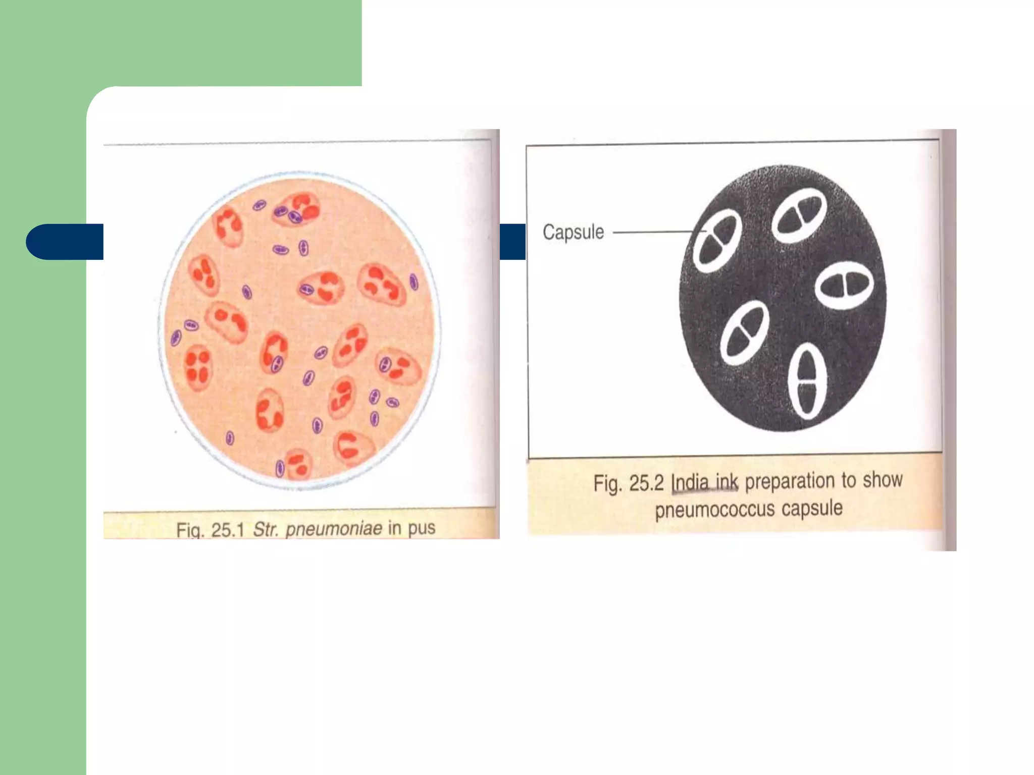
PNEUMOCOCCUS:
Streptococcus pneumoniae
•
•Streptococci
– Gram positive cocci
– chains or pairs
– catalase negative
– facultative anaerobe
(staphylococci are catalase positive)
Streptococcus in chains
groupable streptococci
 A, B and D
– most important
 C, G, F
– rare
hemolysis reaction - sheep blood agar
  (alpha)
– partial hemolysis
 green color
  (beta)
– complete clearing
– A and B
  (gamma)
– no lysis
Classification according to hemolysis
 Groups A an B
– 
 Group D
–  or 
 S. pneumoniae and viridans
– 
PNEUMOCOCCUS:
Streptococcus pneumoniae
Introduction
 Pneumococci are normal commensals of the
upper respiratory tract
 Important pathogen of pneumonia & otitis media
 Reclassified as Streptococcus pneumoniae
 Differ from Streptococci in morphology, bile
solubility, optochin sensitivity & capsule
MORPHOLOGY
 Gram positive cocci
 Flame shaped/lanceolate apperance with
one end broad and the other pointed.
 They are arranged in pairs (diplococci)
 They are capsulated, nonsporing and
nonmotile.
 The capsule is visualised with Indian ink
negative staining.
India-ink preparation Gram stain preparation
Streptococcus pneumoniae
(diplococcus). Fluorescent stain
CULTURAL CHARECTERSTICS
 Pneumococci are fastidious.
 They grows only on enriched media.
 They are aerobic and facultative anaerobic.
 Grows at optimum temperature of 37º C.
 The growth is enhanced by the presence of
5-10% Co2.
 Growth on blood agar:
– Colonies are small, dome shaped.
– With area of green discolouration (alpha
hemolysis) around them.
– On further incubation,
 Concentric rings are seen on the surface when viewed
from above (draughtsman and carrom coin appearance)
Beta hemolytic Alpha haemolytic streptococci
Colonies on blood agar after 18 hrs incubation
Blood agar plate showing draughtsman colonies
of pneumococcus on prolonged incubation
BIOCHEMICAL REACTION
 Catalase negative.
 Fermentation of inulin by Pneumococci is
used for differentiating them from other
streptococci as the later does not ferment it.
 They are bile soluble.
 Optochin sensitive
21
Not optochin sensitive
optochin sensitive
•Most rapid & most useful: identifies & specifies
type of pneumococci in sputum, spinal fluid,
exudates or culture
•Pneumococcal specimen mixed with (polyvalent)
antipneumococcla serum & methylene blue:
•Positive result: Refractile & swollen capsule under oil
immersion
Quellung Reaction
ANTIGENIC STRUCTURE
 The most important antigen of the
Pneumococci is:
– Capsular polysaccharide
– Somatic M protein
– Cell wall cabohydrates(C-substance)
CAPSULAR POLYSACCHRIDES
 This is type specific.
 Pneumococci are classified into types based on the
nature of capsular polysaccharide.
 More than 90 serotypes are recognized.
 The virulence of Pneumococci is dependent upon its
capsule which prevents or inhibits phagocytosis.
 The antibody of capsular polysaccharide protects
against infection.
M Protein
 M Protein is characteristics for each type of
Pneumococci.
 It is not associated with virulence and
antibody to M Protein is non protective.
C- Substance
 Pneumococci contain a species
carbohydrate antigen which is named as C-
Substance.
 It is also detected in blood of patients with
some illness , this is known as C- Reactive
protein (CRP).
TOXINS AND OTHER VIRULENCE
FACTORS.
 Pneumococci produce an:
– Hemolysin.
– Leucocidin
 -Pneumolysin, is a toxin produced by
Pneumococci.
– It has cytolytic and complement activating
properties.
– It is immunogenic.
Pneumococcal disease
Clinical manifestations
 Pneumonia
 Bacteremia
 Meningitis
PATHOGENESIS
 Pneumococcus is one of the most common
bacteria causing pneumonia.
– Lobar pneumonia
– bronchopneumonia
Meningitis
 It is most serious of pneumococcal infection.
It is the second most important cause of
pyogenic meningitis after N.meningitidis.
 Common in children.
 Spreads from the pharynx to the meninges
via blood stream.
Other infections
 Pneumococci may also produce:
– Empyema
– Pericarditis
– Otitis media
– Peritonitis
– suppurative arthritis.
Laboratory Diagnosis
 Samples :according to site
 Collect and transport
 Direct microscopy
 Culture and identification
 Antibiotic sensitivity
Inhibition by optochin
Growth of colonies of Streptococcus
pneumoniae is inhibited by optochin contained in
the disk applied to the blood agar plate
Lysis by bile salts:
Bile salt such as sodium deoxycholate, dissolves
Streptococcus pneumoniae and clear the
turbidity of the heavy inoculum of organisms
Quellung reaction
Capsule of Streptococcus pneumoniae
swells in the presence of specific
pneumococcal antiserum
Morphology
They are gram positive cocci arranged in pairs.
Lanconate shaped
Capsulated organism

Pneumococcus.ppt

  • 1.
  • 2.
    • •Streptococci – Gram positivecocci – chains or pairs – catalase negative – facultative anaerobe (staphylococci are catalase positive)
  • 3.
  • 4.
    groupable streptococci  A,B and D – most important  C, G, F – rare
  • 5.
    hemolysis reaction -sheep blood agar   (alpha) – partial hemolysis  green color   (beta) – complete clearing – A and B   (gamma) – no lysis
  • 6.
    Classification according tohemolysis  Groups A an B –   Group D –  or   S. pneumoniae and viridans – 
  • 7.
  • 8.
    Introduction  Pneumococci arenormal commensals of the upper respiratory tract  Important pathogen of pneumonia & otitis media  Reclassified as Streptococcus pneumoniae  Differ from Streptococci in morphology, bile solubility, optochin sensitivity & capsule
  • 9.
    MORPHOLOGY  Gram positivecocci  Flame shaped/lanceolate apperance with one end broad and the other pointed.  They are arranged in pairs (diplococci)  They are capsulated, nonsporing and nonmotile.  The capsule is visualised with Indian ink negative staining.
  • 10.
    India-ink preparation Gramstain preparation
  • 12.
  • 13.
    CULTURAL CHARECTERSTICS  Pneumococciare fastidious.  They grows only on enriched media.  They are aerobic and facultative anaerobic.  Grows at optimum temperature of 37º C.  The growth is enhanced by the presence of 5-10% Co2.
  • 14.
     Growth onblood agar: – Colonies are small, dome shaped. – With area of green discolouration (alpha hemolysis) around them. – On further incubation,  Concentric rings are seen on the surface when viewed from above (draughtsman and carrom coin appearance)
  • 15.
    Beta hemolytic Alphahaemolytic streptococci
  • 16.
    Colonies on bloodagar after 18 hrs incubation
  • 17.
    Blood agar plateshowing draughtsman colonies of pneumococcus on prolonged incubation
  • 18.
    BIOCHEMICAL REACTION  Catalasenegative.  Fermentation of inulin by Pneumococci is used for differentiating them from other streptococci as the later does not ferment it.  They are bile soluble.  Optochin sensitive
  • 21.
  • 22.
    •Most rapid &most useful: identifies & specifies type of pneumococci in sputum, spinal fluid, exudates or culture •Pneumococcal specimen mixed with (polyvalent) antipneumococcla serum & methylene blue: •Positive result: Refractile & swollen capsule under oil immersion Quellung Reaction
  • 24.
    ANTIGENIC STRUCTURE  Themost important antigen of the Pneumococci is: – Capsular polysaccharide – Somatic M protein – Cell wall cabohydrates(C-substance)
  • 25.
    CAPSULAR POLYSACCHRIDES  Thisis type specific.  Pneumococci are classified into types based on the nature of capsular polysaccharide.  More than 90 serotypes are recognized.  The virulence of Pneumococci is dependent upon its capsule which prevents or inhibits phagocytosis.  The antibody of capsular polysaccharide protects against infection.
  • 26.
    M Protein  MProtein is characteristics for each type of Pneumococci.  It is not associated with virulence and antibody to M Protein is non protective.
  • 27.
    C- Substance  Pneumococcicontain a species carbohydrate antigen which is named as C- Substance.  It is also detected in blood of patients with some illness , this is known as C- Reactive protein (CRP).
  • 28.
    TOXINS AND OTHERVIRULENCE FACTORS.  Pneumococci produce an: – Hemolysin. – Leucocidin  -Pneumolysin, is a toxin produced by Pneumococci. – It has cytolytic and complement activating properties. – It is immunogenic.
  • 29.
    Pneumococcal disease Clinical manifestations Pneumonia  Bacteremia  Meningitis
  • 30.
    PATHOGENESIS  Pneumococcus isone of the most common bacteria causing pneumonia. – Lobar pneumonia – bronchopneumonia
  • 31.
    Meningitis  It ismost serious of pneumococcal infection. It is the second most important cause of pyogenic meningitis after N.meningitidis.  Common in children.  Spreads from the pharynx to the meninges via blood stream.
  • 32.
    Other infections  Pneumococcimay also produce: – Empyema – Pericarditis – Otitis media – Peritonitis – suppurative arthritis.
  • 33.
    Laboratory Diagnosis  Samples:according to site  Collect and transport  Direct microscopy  Culture and identification  Antibiotic sensitivity
  • 34.
    Inhibition by optochin Growthof colonies of Streptococcus pneumoniae is inhibited by optochin contained in the disk applied to the blood agar plate Lysis by bile salts: Bile salt such as sodium deoxycholate, dissolves Streptococcus pneumoniae and clear the turbidity of the heavy inoculum of organisms Quellung reaction Capsule of Streptococcus pneumoniae swells in the presence of specific pneumococcal antiserum Morphology They are gram positive cocci arranged in pairs. Lanconate shaped Capsulated organism