Downloaded 1,450 times


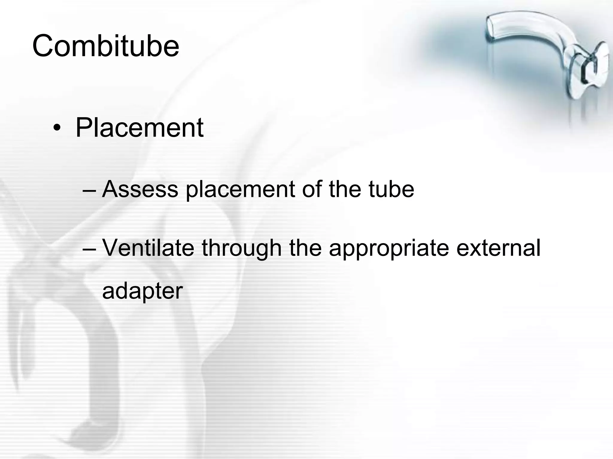


This document discusses various airway management techniques including coughing, suctioning, artificial airways like oropharyngeal and nasopharyngeal tubes, endotracheal tubes, tracheostomy tubes, and alternative devices like LMAs and Combitubes. It provides details on the components of an effective cough, phases of suctioning, indications for different airway techniques, proper procedures, potential hazards, and equipment required.


