Downloaded 530 times


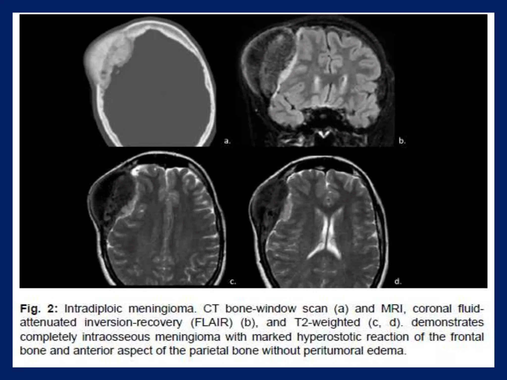


Meningiomas account for 15% of all intracranial tumors and originate from the dura or arachnoid membranes. They are most common in middle-aged adults and affect women twice as often as men. Meningiomas are typically benign, slow-growing tumors that indent the brain as they enlarge. On CT imaging, meningiomas appear well-circumscribed, homogeneous, and hyperdense, and may induce hyperostosis of adjacent bone. MRI often reveals a characteristic "dural tail" sign of enhancement. Other histologic variants include hemangiopericytomas, which have a narrow dural attachment and lobulated shape.


