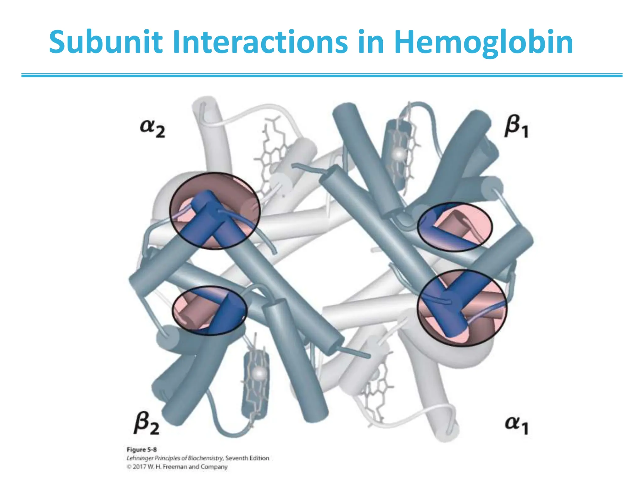
5| Protein Function
© 2017 W. H. Freeman and Company
Learning goals:
• Methods of binding ligands and proteins
• Quantitative and graphical modeling of protein-ligand
interactions
• Interaction of globins with oxygen and non-oxygen
ligands
• Physiological regulation of oxygen binding
• Mechanism and control of antibody-antigen interaction
• Mechanism of muscle contraction
• Regulation of muscle contraction
CHAPTER 5:
Protein Function
Function of Globular Proteins
• Reversible binding of ligands is essential.
– specificity of ligands and binding sites
– Ligand binding is often coupled to conformational
changes, sometimes quite dramatically (induced fit).
– In multisubunit proteins, conformational changes in
one subunit can affect the others (cooperativity).
– Interactions can be regulated.
• Illustrated by:
– hemoglobin, antibodies, and muscle proteins
Key topics in protein function:
Functions of Globular Proteins
• Storage of ions and molecules
– myoglobin, ferritin
• Transport of ions and molecules
– hemoglobin, serotonin transporter
• Defense against pathogens
– antibodies, cytokines
• Muscle contraction
– actin, myosin
• Biological catalysis
– chymotrypsin, lysozyme
Interaction with Other Molecules
• Reversible, transient process of chemical equilibrium:
A + B ↔AB
• A molecule that binds to a protein is called a ligand.
– typically a small molecule
• A region in the protein where the ligand binds is called
the binding site.
• Ligand binds via same noncovalent interactions that
dictate protein structure (see Chapter 4).
– allows the interactions to be transient
Example: Oxygen Binding to Myoglobin
[L]
[L]
d 

K

2
50
2
O
O
p
p
p



When a ligand is a gas, binding is
expressed in terms of partial pressures.
Examples of Binding Strength
Specificity: Lock-and-Key Model
• Proteins typically have high specificity: only certain ligands bind.
• High specificity can be explained by the complementary of the
binding site and the ligand.
• Complementary in:
– size
– shape
– charge
– hydrophobic/hydrophilic character
• The “lock and key” model by Emil Fisher (1894) assumes that
complementary surfaces are preformed.
+
Case Study I: Globins Are Oxygen-
Binding Proteins
Biological problems:
• Protein side chains lack affinity for O2.
• Some transition metals bind O2 well but would generate free
radicals if free in solution.
• Organometallic compounds such as heme are more suitable, but
Fe2+ in free heme could be oxidized to Fe3+ (very reactive!).
Biological solution:
• Capture the oxygen molecule with heme that is protein bound.
Myoglobin (storage) and hemoglobin (transport) can bind oxygen
via a protein-bound heme.
Structures of Porphyrin and Heme
Structure of Myoglobin
Binding of Carbon Monoxide
• CO has similar size and shape to O2; it can fit to the same
binding site.
• CO binds heme over 20,000 times better than O2 because the
carbon in CO has a filled lone electron pair that can be donated
to vacant d-orbitals on the Fe2+.
• The protein pocket decreases affinity for CO, but it still binds
about 250 times better than oxygen.
• CO is highly toxic, as it competes with oxygen. It blocks the
function of myoglobin, hemoglobin, and mitochondrial
cytochromes that are involved in oxidative phosphorylation.
CO vs. O2 Binding to Free Heme
Heme Binding to Protein Affects
CO vs. O2 Binding
Spectroscopic Detection of
Oxygen Binding to Globins
• The heme group is a strong chromophore that absorbs both in
ultraviolet and visible range.
• Ferrous form (Fe2+ ) without oxygen has an intense Soret band
at 429 nm.
• Oxygen binding alters the electronic properties of the heme
and shifts the position of the Soret band to 414 nm.
• Binding of oxygen can be monitored by UV-Vis
spectrophotometry.
• Deoxyhemoglobin (in venous blood) appears purplish in color
and oxyhemoglobin (in arterial blood) is red.
• pO2 in lungs is about 13 kPa: it sure binds oxygen well.
• pO2 in tissues is about 4 kPa: it will not release it!
Would lowering the affinity (P50) of myoglobin to oxygen help?
Could Myoglobin Transport O2?
For Effective Transport
Affinity Must Vary with pO2
How Can Affinity to Oxygen Change?
• It must be a protein with multiple binding sites.
• Binding sites must be able to interact with each other.
• This phenomenon is called cooperativity.
– positive cooperativity
• first binding event increases affinity at remaining sites
• recognized by sigmoidal binding curves
– negative cooperativity
• first binding event reduces affinity at remaining sites
Cooperativity
Hemoglobin Binds Oxygen Cooperatively
• Hemoglobin (Hb) is a tetramer of two subunits (a2b2).
• Each subunit is similar to myoglobin.
Subunit Interactions in Hemoglobin
Subunit Interactions: Details
R and T States of Hemoglobin
• T = tense state
– more interactions, more stable
– lower affinity for O2
• R = relaxed state
– fewer Interactions, more flexible
– higher affinity for O2
• O2 binding triggers a T  R conformational change.
• Conformational change from the T state to the R state
involves breaking ion pairs between the α1-b2
interface.
R and T States of Hemoglobin
Conformational Change Is Triggered
by Oxygen Binding
pH Effect on O2 Binding to Hemoglobin
Does acidity increase or decrease the Kd?
pH Effect on O2 Binding to Hemoglobin
•Actively metabolizing tissues generate H+, lowering the
pH of the blood near the tissues relative to the lungs
(catalyzed by carbonic anhydrase).
CO2 + H2O ↔ HCO3
− + H+
•Hb Affinity for oxygen depends on the pH.
– H+ binds to Hb and stabilizes the T state.
• protonates His146, which then forms a salt bridge with Asp94
• leads to the release of O2 (in the tissues)
•The pH difference between lungs and metabolic tissues
increases efficiency of the O2 transport.
• This is known as the Bohr effect.
Hemoglobin and CO2 Export
• CO2 is produced by metabolism in tissues and must be exported.
• 15–20% of CO2 is exported in the form of a carbamate on the
amino terminal residues of each of the polypeptide subunits.
• Notice:
– The formation of a carbamate yields a proton that can
contribute to the Bohr effect.
– The carbamate forms additional salt bridges, stabilizing the T
state.
2,3-Bisphosphoglycerate Regulates O2 Binding
• Negative heterotropic regulator of Hb function
• Present at mM concentrations in erythrocytes
– produced from an intermediate in glycolysis
• Small negatively charged molecule,
binds to the positively charged
central cavity of Hb
• Stabilizes the T states
2,3-BPG Binds to the Central Cavity of hB
2,3-BPG Allows for O2 Release in the Tissues
and Adaptation to Changes in Altitude
Sickle-Cell Anemia Is Due to
a Mutation in Hemoglobin
• Glu6  Val in the b chain of Hb
• The new Valine side chain can bind
to a different Hb molecule to form a
strand similar to the amyloidgenic
proteins discussed in Chapter 4.
• This sickles the red blood cells.
• Untreated homozygous individuals
generally die in childhood.
• Heterozygous individuals exhibit a
resistance to malaria.
Formation of Hb Strands
in Sickle-Cell Anemia
Cellular Immune System
• Antibodies bind to fragments displayed on the surface of
invading cells.
• Phagocytes: specialized cells that eat invaders
• Macrophages: large phagocytes that ingest bacteria that are
tagged by antibodies
Humoral Immune System
• Vertebrates also fight infections with soluble antibodies
that specifically bind antigens.
– Antigens are substances that stimulate production of
antibodies.
• typically macromolecular in nature
• recognized as foreign by the immune system
• coat proteins of bacteria and viruses
• surface carbohydrates of cells or viruses
– Antibodies are proteins that are produced by B cells and that
specifically bind to antigens.
• Binding will mark the antigen for destruction or interfere with its
function.
• A given antibody will bind to a small region (epitope) of the antigen.
• One antigen can have several epitopes.
Antibodies: Immunoglobulin G
Two heavy chains and two light chains
• composed of constant domains and
variable domains
Light chains: one constant and one
variable domain
Heavy chains: three constant and one
variable domain
Variable domains of each chain make up
the antigen-binding site (two per
antibody) and are hypervariable, which
confers antigen specificity.
Chapter 5: Summary
• how ligand binding can affect protein function
• how to quantitatively analyze binding data
• how myoglobin stores oxygen
• how hemoglobin transports O2, protons, and CO2
• how antibodies recognize foreign structures
• how muscle works
In this chapter, we learned:

Protein Function - General Biology 2 Lesson

  • 1.
    5| Protein Function ©2017 W. H. Freeman and Company
  • 2.
    Learning goals: • Methodsof binding ligands and proteins • Quantitative and graphical modeling of protein-ligand interactions • Interaction of globins with oxygen and non-oxygen ligands • Physiological regulation of oxygen binding • Mechanism and control of antibody-antigen interaction • Mechanism of muscle contraction • Regulation of muscle contraction CHAPTER 5: Protein Function
  • 3.
    Function of GlobularProteins • Reversible binding of ligands is essential. – specificity of ligands and binding sites – Ligand binding is often coupled to conformational changes, sometimes quite dramatically (induced fit). – In multisubunit proteins, conformational changes in one subunit can affect the others (cooperativity). – Interactions can be regulated. • Illustrated by: – hemoglobin, antibodies, and muscle proteins Key topics in protein function:
  • 4.
    Functions of GlobularProteins • Storage of ions and molecules – myoglobin, ferritin • Transport of ions and molecules – hemoglobin, serotonin transporter • Defense against pathogens – antibodies, cytokines • Muscle contraction – actin, myosin • Biological catalysis – chymotrypsin, lysozyme
  • 5.
    Interaction with OtherMolecules • Reversible, transient process of chemical equilibrium: A + B ↔AB • A molecule that binds to a protein is called a ligand. – typically a small molecule • A region in the protein where the ligand binds is called the binding site. • Ligand binds via same noncovalent interactions that dictate protein structure (see Chapter 4). – allows the interactions to be transient
  • 6.
    Example: Oxygen Bindingto Myoglobin [L] [L] d   K  2 50 2 O O p p p    When a ligand is a gas, binding is expressed in terms of partial pressures.
  • 7.
  • 8.
    Specificity: Lock-and-Key Model •Proteins typically have high specificity: only certain ligands bind. • High specificity can be explained by the complementary of the binding site and the ligand. • Complementary in: – size – shape – charge – hydrophobic/hydrophilic character • The “lock and key” model by Emil Fisher (1894) assumes that complementary surfaces are preformed. +
  • 9.
    Case Study I:Globins Are Oxygen- Binding Proteins Biological problems: • Protein side chains lack affinity for O2. • Some transition metals bind O2 well but would generate free radicals if free in solution. • Organometallic compounds such as heme are more suitable, but Fe2+ in free heme could be oxidized to Fe3+ (very reactive!). Biological solution: • Capture the oxygen molecule with heme that is protein bound. Myoglobin (storage) and hemoglobin (transport) can bind oxygen via a protein-bound heme.
  • 10.
  • 11.
  • 12.
    Binding of CarbonMonoxide • CO has similar size and shape to O2; it can fit to the same binding site. • CO binds heme over 20,000 times better than O2 because the carbon in CO has a filled lone electron pair that can be donated to vacant d-orbitals on the Fe2+. • The protein pocket decreases affinity for CO, but it still binds about 250 times better than oxygen. • CO is highly toxic, as it competes with oxygen. It blocks the function of myoglobin, hemoglobin, and mitochondrial cytochromes that are involved in oxidative phosphorylation.
  • 13.
    CO vs. O2Binding to Free Heme
  • 14.
    Heme Binding toProtein Affects CO vs. O2 Binding
  • 15.
    Spectroscopic Detection of OxygenBinding to Globins • The heme group is a strong chromophore that absorbs both in ultraviolet and visible range. • Ferrous form (Fe2+ ) without oxygen has an intense Soret band at 429 nm. • Oxygen binding alters the electronic properties of the heme and shifts the position of the Soret band to 414 nm. • Binding of oxygen can be monitored by UV-Vis spectrophotometry. • Deoxyhemoglobin (in venous blood) appears purplish in color and oxyhemoglobin (in arterial blood) is red.
  • 16.
    • pO2 inlungs is about 13 kPa: it sure binds oxygen well. • pO2 in tissues is about 4 kPa: it will not release it! Would lowering the affinity (P50) of myoglobin to oxygen help? Could Myoglobin Transport O2?
  • 17.
  • 18.
    How Can Affinityto Oxygen Change? • It must be a protein with multiple binding sites. • Binding sites must be able to interact with each other. • This phenomenon is called cooperativity. – positive cooperativity • first binding event increases affinity at remaining sites • recognized by sigmoidal binding curves – negative cooperativity • first binding event reduces affinity at remaining sites
  • 19.
  • 20.
    Hemoglobin Binds OxygenCooperatively • Hemoglobin (Hb) is a tetramer of two subunits (a2b2). • Each subunit is similar to myoglobin.
  • 21.
  • 22.
  • 23.
  • 24.
    R and TStates of Hemoglobin • T = tense state – more interactions, more stable – lower affinity for O2 • R = relaxed state – fewer Interactions, more flexible – higher affinity for O2 • O2 binding triggers a T  R conformational change. • Conformational change from the T state to the R state involves breaking ion pairs between the α1-b2 interface.
  • 25.
    R and TStates of Hemoglobin
  • 26.
    Conformational Change IsTriggered by Oxygen Binding
  • 27.
    pH Effect onO2 Binding to Hemoglobin Does acidity increase or decrease the Kd?
  • 28.
    pH Effect onO2 Binding to Hemoglobin •Actively metabolizing tissues generate H+, lowering the pH of the blood near the tissues relative to the lungs (catalyzed by carbonic anhydrase). CO2 + H2O ↔ HCO3 − + H+ •Hb Affinity for oxygen depends on the pH. – H+ binds to Hb and stabilizes the T state. • protonates His146, which then forms a salt bridge with Asp94 • leads to the release of O2 (in the tissues) •The pH difference between lungs and metabolic tissues increases efficiency of the O2 transport. • This is known as the Bohr effect.
  • 29.
    Hemoglobin and CO2Export • CO2 is produced by metabolism in tissues and must be exported. • 15–20% of CO2 is exported in the form of a carbamate on the amino terminal residues of each of the polypeptide subunits. • Notice: – The formation of a carbamate yields a proton that can contribute to the Bohr effect. – The carbamate forms additional salt bridges, stabilizing the T state.
  • 30.
    2,3-Bisphosphoglycerate Regulates O2Binding • Negative heterotropic regulator of Hb function • Present at mM concentrations in erythrocytes – produced from an intermediate in glycolysis • Small negatively charged molecule, binds to the positively charged central cavity of Hb • Stabilizes the T states
  • 31.
    2,3-BPG Binds tothe Central Cavity of hB
  • 32.
    2,3-BPG Allows forO2 Release in the Tissues and Adaptation to Changes in Altitude
  • 33.
    Sickle-Cell Anemia IsDue to a Mutation in Hemoglobin • Glu6  Val in the b chain of Hb • The new Valine side chain can bind to a different Hb molecule to form a strand similar to the amyloidgenic proteins discussed in Chapter 4. • This sickles the red blood cells. • Untreated homozygous individuals generally die in childhood. • Heterozygous individuals exhibit a resistance to malaria.
  • 34.
    Formation of HbStrands in Sickle-Cell Anemia
  • 35.
    Cellular Immune System •Antibodies bind to fragments displayed on the surface of invading cells. • Phagocytes: specialized cells that eat invaders • Macrophages: large phagocytes that ingest bacteria that are tagged by antibodies
  • 36.
    Humoral Immune System •Vertebrates also fight infections with soluble antibodies that specifically bind antigens. – Antigens are substances that stimulate production of antibodies. • typically macromolecular in nature • recognized as foreign by the immune system • coat proteins of bacteria and viruses • surface carbohydrates of cells or viruses – Antibodies are proteins that are produced by B cells and that specifically bind to antigens. • Binding will mark the antigen for destruction or interfere with its function. • A given antibody will bind to a small region (epitope) of the antigen. • One antigen can have several epitopes.
  • 37.
    Antibodies: Immunoglobulin G Twoheavy chains and two light chains • composed of constant domains and variable domains Light chains: one constant and one variable domain Heavy chains: three constant and one variable domain Variable domains of each chain make up the antigen-binding site (two per antibody) and are hypervariable, which confers antigen specificity.
  • 38.
    Chapter 5: Summary •how ligand binding can affect protein function • how to quantitatively analyze binding data • how myoglobin stores oxygen • how hemoglobin transports O2, protons, and CO2 • how antibodies recognize foreign structures • how muscle works In this chapter, we learned: