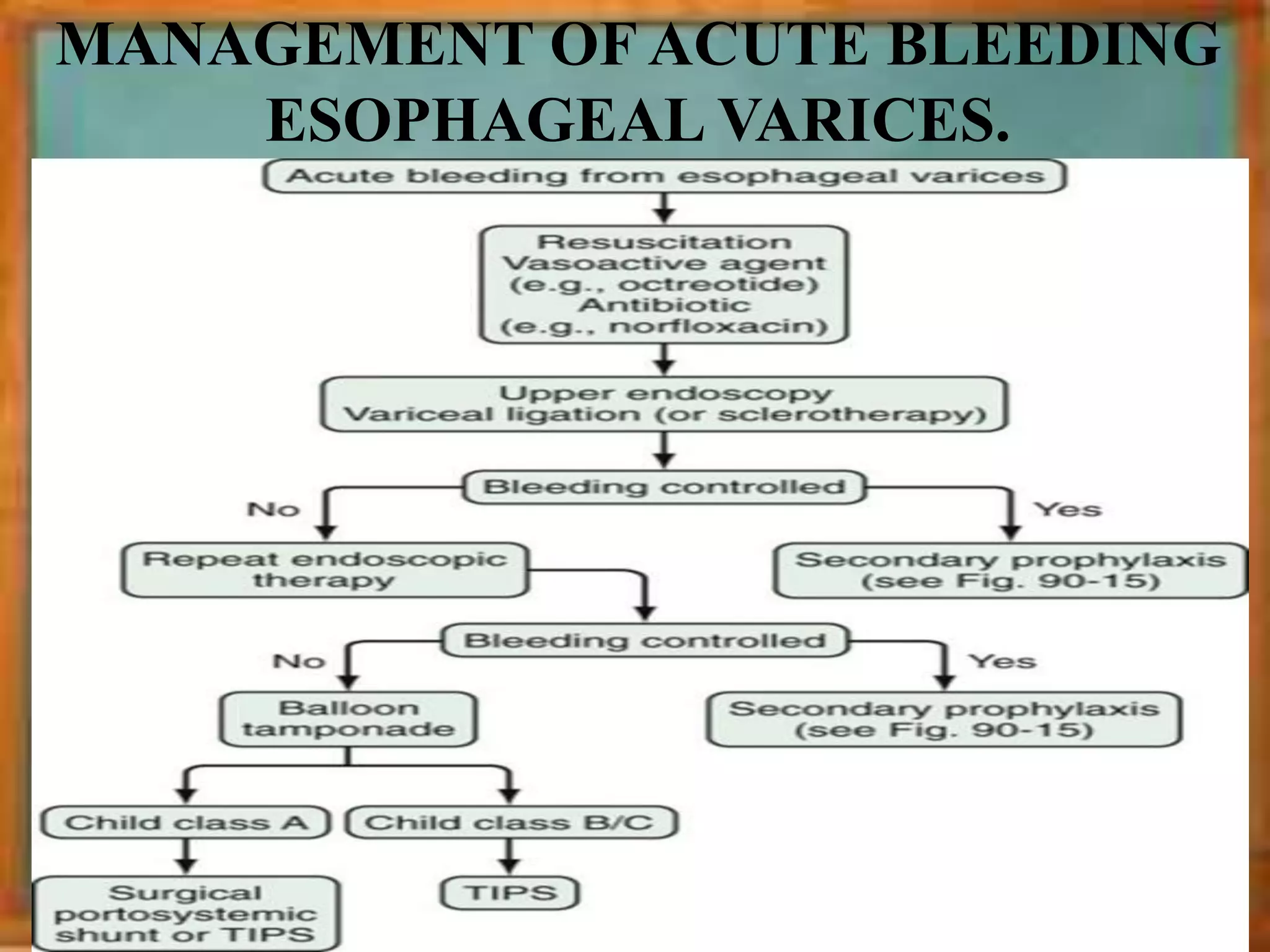
APPROACH TO UPPER GI BLEEDING
By :- Dr. NITESH PANSARI
MEDICINE UNIT –A
RNT MEDICAL COLLEGE , UDAIPUR
SYNOPSIS
• Definition & Presentation
• Causes of UGI bleeding
• History & Physical Examination
• Investigations
• Management of acute episode
• Variceal bleeeding & management
• Nonvariceal bleeding & management
• High risk Factors
• Scoring systems
• Indication of surgical intervention
DEFINITION
• Upper GI bleeding is defined as bleeding from a
source proximal to the Ligament of Treitz
Hematemesis
• Vomiting of red blood or coffee- grounds material
when gastric acid converts hemoglobin into
methemoglobin .
• Differentiate from :-
Hemoptysis.
Bleeding from Pharynx , nasal passage.
Patient with hematemesis usually requires a bucket;
whereas patient with hemoptysis usually requires a small
bowl.
Is It Hematemesis?
Melena
• Passage of black tarry stools.
• Blood loss between 50-100 ml /day will produce
melena.
• The black color of melenic stools is caused by
Hematin, the product of oxidation of Heme by
intestinal and bacterial enzymes.
• Can be seen in LGI bleed also (10%).
• Non GI bleed – swallowed blood from epistaxis
• Blood for 14 hrs in the GI tract
• Oral iron and bismuth mimics melena.
Hematochezia
• It is defined as passage of bright-red or maroon blood from the rectum.
• Common in bleeding from colon, rectum and anus.
• In case of brisk bleeding in the upper git, bright red blood may come out
unchanged in the stool.
• 10% of UGI bleed
Occult GI bleeding
• Absence of overt bleeding
• Fecal occult blood test - +ve
• Suspected in patients with iron deficiency anemia
• Normally 2.5 ml of blood is lost per day.
• OBT detects amount between 10-50 ml/d.
Symptoms of blood loss or anemia
• Light headedness, syncope, angina, or dyspnea
UPPER GI
BLEEDING
VARICEAL
BLEEDING
NON VARICEAL
BLEEDING
Esophageal causes
• Esophageal varices
• Esophagitis
• Esophageal cancer
• Esophageal ulcers
• Mallory-Weiss tear
Gastric causes
• Gastric ulcer
• Gastric cancer
• Gastritis
• Gastric varices
• Dieulafoy's lesions
• Gastric Antral
Vascular Ectasia
• Portal Hypertensive
Gastropathy
Duodenal causes
• Duodenal ulcer
• Vascular
malformation
including aorto-
enteric fistulae
• Hematobilia, or
bleeding from the
biliary tree
• Hemosuccus
pancreaticus, or
bleeding from the
pancreatic duct
• Severe superior
mesenteric artery
syndrome
CAUSES
Bleeding etiology Leading History
Mallory -Weiss tear Multiple Emesis before hematemesis, alcoholism, retching
Esophageal ulcer Dysphagia, Odynophagia, GERD
Peptic ulcer Epigastric pain, NSAID or aspirin use
Stress gastritis Patient in an ICU, gastrointestinal bleeding occurring
after admission,respiratory failure,multiorgan failure,coagulopathy
Varices, portal
gastropathy
Alcoholism, Cirrhosis of liver
Gastric antral
vascular ectasia
Renal failure, cirrhosis
Malignancy Recent involuntary weight loss, dysphagia, cachexia,
early satiety
Angiodysplasia Chronic renal failure, hereditary hemorrhagic
telangiectasia
Aortoenteric fistula Known aortic aneurysm, prior abdominal aortic
aneurysm repair
Clues regarding the cause of acute UGI bleeding
• Previous Episodes Of GI Bleed
• H/O Instrumentation
• H/O Head Injury
• Co-morbid Conditions
o Liver Disease
o Renal Disease
o Cardiovascular Disease
o Chronic Respiratory Conditions
• H/O Abdominal Surgery
Personal History
• Alcoholism , Smoking , Tobacco Abuse
• Cocaine Abuse
• Weight Loss, Anorexia
Past History
Drug History
• Salicylates/ NSAIDS
• Anticoagulants (warfarin / Heparin)
• Corticosteroids
• Oral Iron , Bismuth ( Mimic Melena )
• Pallor , signs of dehydration , Shock
• Icterus
• Clubbing
• Oedema
}Liver disease
PHYSICAL EXAMINATION
Lymphadenopathy
Virchow’s Node
(Troisier’s sign)
Vital signs
Tachycardia
Hypotension
Tachypnea
SIGNS OF LIVER CELL
FAILURE
• Ascites • Spider naevi
• Palmar erythema • Dupuytren’s contracture
• Leuconychia • Gynecomastia
• Bleeding manifestations • Scars of previous surgery
• Splenomegaly
• Caput medusae
• Parotid Swelling
• Fetor hepaticus
• Asterixis
• Testicular atrophy
• Acanthosis nigricans
• Alopecia
• Glossitis
• Loss of Axillary hair
• Loss of Pubic Hair
INSPECTION
Distention , Dilated Veins
Swelling
Visible peristalsis
PALPATION
Tenderness
HSM, secondary metastasis
Sister Mary Joseph nodules
Mass
PERCUSSION
Shifting dullness
HSM
AUSCULTATION
Absent Bowel sound
Bruit
Cruveilhier-Baumgarten venous hum
ABDOMEN EXAMINATION
PR EXAMINATION
• Hemorroids
• Melena , blood
• Blumer’s shelf
LAB ASSESSMENT
• Full Blood Count- Hb, Platelet count
• PCV*
• Coagulation Profile
• Liver Function tests
• Serum electrolytes
• Blood urea nitrogen ratio > 30 sensitivity of 68% and a
specificity of 98%
• Blood Grouping & Cross matching.
• Serial ECG
• Stool Occult Blood Test
• HIV, HbsAg , Anti HCV Markers
INVESTIGATIONS
• Renal Function Tests
• Gastrin level
• Nasogastric aspiration
1. Red blood-current bleeding
2. Coffee ground-recent bleeding
3. Continuous aspiration-severe active bleed
Lavage not +ve- i) bleeding has stopped
ii) beyond pylorus
• PCV* : decreased only by 24 to 72hrs, after
bleed
 CHEST XRAY
 USG
ESOPHAGOGASTRODUODENOSCOPY
• Most important component of investigation.
• 90% accuracy in diagnosis if done with in 24 hours.
ENDOSCOPY
•Examination of whole bowel possible
•Indicated in GI bleeding of obscure source
Video capsule endoscopy
•PUSH ENTEROSCOPY
•DEEP ENTEROSCOPY
 SINGLE OR DOUBLE
BALLOON ENDOSCOPY
 SPIRAL ENDOSCOPY
RADIOLOGIC IMAGING
• ANGIOGRAPHY :- bleeding rate at least
0.5 mL/min.
• RADIONUCLIDE IMAGING :- bleeding at a rate
of 0.04 mL/min
Technetium pertechnetate–labeled red blood
cell scans >>> Technetium sulfur colloid scans
• CT enterography and CT angiography
• Barium radiography is not indicated (and
contraindicated if endoscopy or angiography is
planned) in the emergency setting.
Airway:
• Secure to prevent aspiration
• Endotracheal tube & give oxygen
Breathing:
• support respiratory function
Circulation:
• Expand circulatory volume
• 2 large bore i.v. cannula
• RBC transfusion with the goal of maintaining the
hematocrit value to 30% or hemoglobin to 8 g/dL.
• Normal saline may be infused intravenously until packed
red blood cells are available for transfusion.
• Maintain systolic B.P. at equal or greater than 90mmHg
• Pressure agents like dopamine 2.5 to 10 mcg/kg/min, and
dobutamine 2.5 to 10 mcg/kg/min as infusion,
Noradrenalin 3mg. /h as infusion may be required, as a
last resort, to maintain peripheral perfusion.
• Correct any coagulopathy with :-
10 - 15 ml/kg of FFP (if PT INR > 1.5) and/or
platelet transfusions (if platelet count < 50,000/cu.mm)
• Monitor:
 Skin color, peripheral temperature
 Pulse Rate, BP
 Respiratory Rate, O2 saturation of blood
 Urine output (Foley’s Catheter)
TREATMENT OF SUSPECTED PEPTIC ULCER
BLEEDING:
• Start High dose , constant infusion IV PPIs (Omeprazole 80 mg
bolus and 8mg/ h infusion for 72 h)
TREATMENT OF SUSPECTED
VARICEAL HEMORRHAGE
 Antibiotic prophylaxis
• To prevent bacteremia and spontaneous bacterial peritonitis
• Intravenous ceftriaxone, 1 g every 24 hours for 7 days ; or
• Norfloxacin, 400 mg orally twice daily for 7 days, is the preferred choice.
• Intravenous ciprofloxacin, 400 mg every 12 hours; or
• Intravenous levofloxacin, 500 mg every 24 hours
 Pharmacological therapy
• Pharmacological therapy (somatostatin or its analogues octreotide and
vapreotide; terlipressin) should be initiated and continued for 3-5 days after
diagnosis is confirmed.
 Inj. Vit K.
 Correct Hepatic encephalopathy by lactulose and phosphate enemas.
 Avoid Sedatives.
 If tense ascites is present , paracentesis may be done and iv albumin and
spironolactone may be used .
VARICEAL BLEEDING
AND MANAGEMENT
VARICEAL
BLEEDING
ESOPHAGEAL
VARICES
GASTRIC
VARICES
ECTOPIC
VARICES
ESOPHAGEAL VARICES
• Present in approximately 40% of patients with
cirrhosis and in 60% of patients with cirrhosis and
ascites.
• Up to 25% of patients with newly diagnosed varices
will bleed within two years.
• Approximately one half of patients with a variceal
bleed stop bleeding spontaneously.
• The risk of bleeding in patients with varices:-
 less than 5 mm in diameter is 7% by two years.
 greater than 5 mm in diameter is 30% by two years.
• The risk of death with acute variceal bleeding is 5%
to 8% at one week and about 20% at six weeks.
GRADING OF ESOPHAGEAL VARICES
Japanese Research Society for
Portal Hypertension
• Esophageal varices may be :-
• Grade I: small and straight
• Grade II : tortuous and occupying
less than one third of the
esophageal lumen
• Grade III : large and occupying
more than one third of the
esophageal lumen
Risk of bleeding is higher with
a) Large varices – Grade III > Grade I / II
b) Red Wale sign
c) Child Pugh Turcotte Score – C>B>A
PRIMARY PROPHYLAXIS OF ESOPHAGEAL
VARICEAL HEMORRHAGE
• INDICATIONS:-
 All Patients with Class C cirrhosis who have small varices.
 All patients with large varices (diameter greater than 5 mm).
• In patients who do not bleed during therapy and who do not experience side
effects, treatment should be continued indefinitely.
• The goal of treatment is to reduce the HVPG to <12 mm Hg or by ≥20%.
• The usual starting dose of -
 Long-acting Propranolol is 60 mg once daily (Start as 20 mg BD).
 Nadolol is 20 mg once daily..
• The dose of propranolol or nadolol can be increased gradually every three to
five days until the target heart rate of 25% below baseline or 55 to 60 beats per
minute or the maximum tolerated dose is reached, provided that the systolic
blood pressure remains above 90 mm Hg.
• In patients on pharmacologic therapy, follow-up endoscopy is unnecessary
unless gastrointestinal bleeding occurs.
MANAGEMENT OF ACUTE BLEEDING
ESOPHAGEAL VARICES.
VASOACTIVE AGENTS
• Vasopressin with Nitroglycerin 50-400 µg/min
• Terlipressin( triglycyl-lysine-vasopressin) : 2mg
iv every 6 h for 48 h ; may be continued 1 mg
every 4-6 h for further 3 days.
• Somatostatin : 250 or 500 µg iv bolus f/b
infusion of 6 mg/24 h for 120 h.
• Somatostatin analogs :
• Octreotide 50 µg iv bolus & 50µg /h infusion
for 2-5 days.
• Lanreotide and vapreotide.
• Pharmacologic treatment should be continued
for up to five days to prevent early rebleeding.
BALL00N TAMPONADE
Sengstaken Blakemore Tube
Minnesota four-lumen tube
• Balloon tamponade should be used as a temporizing
measure (maximum 24 hours) in patients with
uncontrollable bleeding for whom a more definitive
therapy (e.g., TIPS or endoscopic therapy) is planned.
ENDOSCOPIC METHODS
ENDOSCOPY
METHODS
ENDOSCOPIC
VARICEAL
LIGATION
ENDOSCOPIC
SCLEROTHERAPY
Inj.
CYANOACRYLATE
GLUE
OGD, performed within 12 hours, should be used to make the
diagnosis and to treat variceal hemorrhage, either with EVL or
sclerotherapy.
ENDOSCOPIC VARICEAL LIGATION (EVL)
• Mechanical strangulation of varices by a rubber band.
• Multiple bands are applied.
• Fall away in 1 to 10 days.
• Sloughing produces ulcers which are shallow and heal
faster as compared to EST.
• Frequency of application of EVL
 Varies from 2wks to 2months.
 The preferred interval is 4wks.
• Complications :
 Esophageal ulcers, laceration, perforation.
 Transient dysphagia, chest pain.
 Bacteremia (EST>10times EVL)
ENDOSCOPIC SCLEROTHERAPY (EST)
• Induces inflammation and thrombosis of varix which leads to
fibrosis and obliteration of varices.
• Used where poor visualisation of varices is present.
SOLUTION FOR EST
 Polidocanol 1-3%
 Sodium morrhuate
 1% Sodium Tetradocyl sulphate
 Absolute alcohol
 5% Ethanolamine oleate
COMPLICATIONS
 Post esophageal ulcers
 Bacteremia – seen in 35%.
 Esophageal stenosis – 2 -10%
 Esophageal perforation – rare. Can occur early or late
 Mortality – 2%. Due to recurrent bleed, perforation, sepsis
REBLEED
• Approximately 33% will rebleed within the
next 6 weeks.
• Of all rebleeding episodes, approximately
40% will take place within 5 days of the
initial bleed.
• Predictors of rebleeding include :-
active bleeding at emergency endoscopy,
bleeding from gastric varices,
hypoalbuminemia,
renal insufficiency,
HVPG greater than 20 mm Hg
Prevention of recurrent variceal bleeding
(secondary prophylaxis)
• Combined therapy with endoscopic variceal
ligation and a nonselective beta blocker is the
preferred treatment.
• If goals are not achieved, isosorbide mononitrate
may be added.
• Isosorbide mononitrate-an initial starting dose of
30 mg/day.
• Patients with a CTP score of 7 or greater also
should be evaluated for liver transplantation.
TRANSJUGULAR INTRAHEPATIC
PORTOSYSTEMIC SHUNT (TIPS)
• TIPS is indicated in patients in whom hemorrhage from esophageal
varices cannot be controlled or in whom bleeding recurs despite
combined pharmacological and endoscopic therapy.
• Does not prolong the survival of patients with variceal bleeding.
GASTRIC VARICES
• In contrast with esophageal
varices, bleeding from gastric
varices has been described with
an HVPG less than 12 mm Hg.
• If endoscopic and pharmacologic
therapies fail to control gastric
variceal bleeding, then a Linton-
Nachlas tube, which has a 600-
mL balloon, may be passed as a
temporizing measure.
• Occurs with splenic vein
thrombosis and pancreatitis or
pancreatic cancer.
SARIN CLASSIFICATION
 Type 1 : Gastroesophageal varices (GOV1) extend 2 to 5
cm below the gastroesophageal junction and are in
continuity with esophageal varices;
Type 2 : Gastroesophageal varices (GOV2) are in the
cardia and fundus of the stomach and in continuity with
esophageal varices;
Type 3 : Isolated gastric varices type 1 (IGV1) that occur
in the fundus of the stomach in the absence of esophageal
varices;
Type 4 : Isolated gastric varices type 2 (IGV2) that occur
in the gastric body, antrum , or pylorus.
SARIN CLASSIFICATION
Management of bleeding gastric varices
Management of bleeding from ectopic varices
Snake skin appearance
• One fourth of all cases of
bleeding (acute and chronic)
in patients with portal
hypertension .
• The more common
presentation is one of
chronic, slow bleeding and
anemia.
• A characteristic mosaic-like
pattern of the gastric mucosa.
• Development of PHG
correlates with the duration
of cirrhosis but not
necessarily the degree of liver
dysfunction.
PORTAL HYPERTENSIVE GASTROPATHY
Management Of Chronic Bleeding From Portal Hypertensive
Gastropathy
NON VARICEAL
BLEEDING AND
MANAGEMENT
• Most frequent cause of upper GI Bleed (31-
67% of all cases)
Duodenal Ulcer-gastroduodenal A.
PUD
Gastric ulcer-left gastric A.
PEPTIC ULCER DISEASE
Modified Johnson classification for
gastric ulcer
– Ia: Active Spurting Bleeding
– Ib: Non spurting Active oozing
– IIa: Non bleeding visible vessel
(NBVV)
– IIb: Non bleeding ulcer with
adherent clot
– IIc: Ulcer with hematin
covered base(Flat pigment)
– III: Clean base ulcer ground
Minor
SRH
Major
SRH
62/81
FORREST CLASSIFICATION
FOR BLEEDING PEPTIC ULCER
ENDOSCOPIC
CHARACTERISTIC
FREQUENCY (%)
FURTHER
BLEEDING (%)
SURGERY (%) MORTALITY (%)
Clean base (type
III)
42 5 0.5 2
Flat pigmentation
(type IIc)
20 10 6 3
Adherent clot
(type IIb)
17 22 10 7
Nonbleeding
visible vessel
(type IIa)
17 43 34 11
Active bleeding
(type I)
18 55 35 11
Modalities for endoscopic therapy
Two sessions of endoscopic hemostasis
Angiographic embolization Surgical therapy
PUD (GU OR DU)
H. Pylori
Related
NSAID
Related
If symptoms remain , refer to
Gastroenterologist
After 4 weeks Eradication Test
Triple therapy for 14 days + H2
blockers or PPI for 4-6 weeks
Active
Ulcer
NSAID Discontinued →
H2 blocker Or PPI
NSAID Continued→ PPI
Prophylaxis
Misoprostol
PPI
COX 2
Inhibitor
• H.Pylori Treatment- TRIPLE THERAPY
•
30mgBD 500mgBD
1000 mg BD
• Mallory-Weiss syndrome refers to bleeding from tears (a
Mallory-Weiss tear) in the mucosa at the junction of the
stomach and esophagus, usually caused by severe retching,
coughing, or vomiting.
• Mallory-Weiss tears account for 5% to 10% of cases
Mallory-Weiss Tears
Mallory-weiss tears
• Supportive therapy:-
90% episodes are self limiting.
Mucosa heals within 72hrs.
• Ongoing bleeding-local
endoscopic therapy
• Appearance of multiple superficial erosions of the
entire stomach, most commonly in the body.
 Respiratory failure
 Coagulopathy
 NSAID users
 Sepsis
 Hemodynamic instability
 Head injuries with I.C.T (Cushing ulcer)
 Burn injuries (Curling ulcer)
 Multiple trauma
Stress
Gastritis
decrease
splanchnic
mucosal
blood flow
altered
gastric
luminal
acidity
STRESS GASTRITIS
PROPHYLACTIC THERAPY
•Routine uses of stress ulcer prophylaxis in the
intensive care unit is not recommended unless the
patient has a coagulopathy or is receiving mechanical
ventilation.
•Maintain the luminal gastric pH above 4.
PPIS,,, H2 Antagonists.. Sucralfate..
• PERSISTENT
CALIBER ARTERY
• Vascular malformations
along the lesser curve of
the stomach within 6
cm of the GEJ
• Represents rupture of
unusually large vessels
(1-3 mm) in the gastric
submucosa.
• Erosion of the gastric
mucosa overlying these
vessels leads to
hemorrhage.
• Endoscopic therapy
DIEULAFOY’S LESION
• “Watermelon stomach”
• Collection of dilated
venules appearing as
linear red streaks
converging on the antrum
in longitudinal fashion,
giving it the appearance
of a watermelon.
• GAVE is related to liver
failure, rather than to
portal hypertension.
GASTRIC ANTRAL VASCULAR
ECTASIA
Management of chronic bleeding from gastric vascular antral ectasia
(GAVE)
• Usually associated with
chronic anemia or
hemoccult-positive stool
• Occasionally, malignancies
present as ulcerative lesions
that bleed persistently.
• Most characteristic of the
GIST
• Also occur with leiomyomas
and lymphomas.
MALIGNANCY
Aortoenteric fistula
• Primary aortoenteric fistula is
a communication between the
abdominal aorta and most
commonly, the third portion
of the duodenum.
• Secondary aortoenteric
fistulas usually occur between
the small intestine and an
infected abdominal aortic
surgical graft.
• CT with intravenous contrast
• Surgical treatment
• Typically associated
with liver trauma, liver
biopsy, recent
instrumentation of the
biliary tree, or hepatic
neoplasms.
• Suspected in anyone
who presents with
hemorrhage, right upper
quadrant pain, and
jaundice
• Angiographic
embolisation
HAEMOBILIA
• Bleeding from the pancreatic
duct.
• In patients with acute
pancreatitis, chronic
pancreatitis, pancreatic
pseudocyst, or pancreatic
cancer or after ERCP
• Also result from rupture of a
splenic artery aneurysm into
the pancreatic duct.
• Presents with abdominal pain
and hematochezia
• Angiographic embolization or
Distal pancreatectomy
Haemosuccus Pancreaticus
• May follow therapeutic or diagnostic procedures.
• Common causes of iatrogenic bleeding –
Endoscopic sphincterotomy.
Percutaneous transhepatic procedures.
• 2% of cases.
• It is often mild and self-limited.
• Percutaneous endoscopic gastrotomy.
IATROGENIC BLEEDING
1. Advanced AGE >65 years
2. SHOCK on admission(pulse rate >100 beats/min; systolic
blood pressure < 100mmHg)
3. COMORBIDITY (particularly hepatic or renal failure , DM ,
HTN and disseminated malignancy)
4. Diagnosis (worst PROGNOSIS for advanced upper
gastrointestinal malignancy)
5. ENDOSCOPIC FINDINGS (active, spurting hemorrhage from
peptic ulcer; non-bleeding visible vessel)
6. RECURRENT BLEEDING (increases mortality 10 times)
7. Where there is simultaneous upper and lower GI bleeding.
8. On steroids or NSAIDs
9. Alcoholic or tobacco smoker 80/81
UPPER GI BLEED:
HIGH RISK FACTORS
SCORING SYSTEM
1. Glasgow-Blatchford Score
2. AIMS 65
3. THE ROCKALL SCORE
 Blood Urea Nitrogen(mmol/L)
6.5 – 7.9 2
8 – 9.9 3
10 – 24.9 4
≥25 6
 Haemoglobin (g/dL) for men
12-12.9 1
10-11.9 3
<10 6
 Haemoglobin (g/dL) for
women
10-11.9 1
<10 6
 Systolic BP (mm Hg)
◦ 100–109 1
◦ 90–99 2
◦ <90 3
•Other markers
Pulse ≥100 (per min) 1
Presentation with melena 1
Presentation with syncope 2
Hepatic disease 2
Cardiac failure 2
•Score from 0 to 23
•scores ≥ 6 - 50% risk of needing an
intervention.
Score
Score is"0" if :
•Hemoglobin level
>12.9 g/dl(men) or
>11.9 g/dl(women)
•Systolic blood pressure >109 mm Hg
•Pulse <100/minute
•BUN level <6.5 mmol/L
•No melena or syncope
•No liver disease or heart failure
AIMS 65
High accuracy for predicting inpatient mortality among patients
with upper GI bleeding.
• Albumin less than 3.0 g/dL (30 g/L)
• INR greater than 1.5
• Altered Mental status (Glasgow coma score less than 14, disorientation,
lethargy, stupor, or coma)
• Systolic blood pressure of 90 mmHg or less
• Age older than 65 years
The mortality rate increased significantly as the number of risk
factors present increased:
●Zero risk factors: 0.3 percent
●One risk factor: 1 percent
●Two risk factors: 3 percent
●Three risk factors: 9 percent
●Four risk factors: 15 percent
●Five risk factors: 25 percent
THE ROCKALL SCORE
Rockall, Lancet 1996
• For Risk of Rebleeding and Death After Admission to the
Hospital for Acute GI Bleeding
Risk category
Rockall’s score>8=High risk of death
50% Patients will rebleed.
Rockall’s score<3=excellent prognosis
Lower risk of hemorrhage.
Rockall score
Cumulative
patients
with
rebleeding
Who can be sent home from the emergency room?
Gralnek, NEJM, 2008
These patients
represent up
to 20-40% of
all patients
presenting with
PUB
SURGICAL MANAGEMENT
• Hemodynamic instability
despite vigorous resuscitation
(>6 units transfusion)
• Failure of endoscopy
• Recurrent hemorrhage after
initial stabilization (with up
to two attempts of
endoscopy)
• Shock associated with
recurrent hemorrhage
• Continued slow bleeding
with a transfusion exceeding
3 units/day
 Oneofthecriteriausedtodeterminetheneedforsurgicalinterventionis
thenumberofunitsoftransfusedbloodrequiredtoresuscitatethepatient.
Themoreunitsrequired,thehigherthemortalityrate(Larson,1986).
Operativeinterventionisindicatedoncethebloodtransfusionnumber
reachesmorethan5units,asnotedinthefollowingtable(Larson,1986).
NumberofUnits
Transfused
Needfor
Surgery,%
Mortality
Rate,%
0 4 4
1-3 6 14
4-5 17 28
>5 57 43
Indications for Surgery in Gastrointestinal
Hemorrhage :-
Relative Indications
• Rare blood type or difficult crossmatch
• Refusal to transfusion
• Shock at presentation
• Advanced age
• Severe comorbid diseases
• Bleeding chronic gastric ulcer where
malignancy is a possibility
PRIORITIES
•Control Hemorrhage !!
• Definitive procedure for the underlying pathology
uppergibleeding-150402032909-conversion-gate01.pdf

uppergibleeding-150402032909-conversion-gate01.pdf

  • 1.
    APPROACH TO UPPERGI BLEEDING By :- Dr. NITESH PANSARI MEDICINE UNIT –A RNT MEDICAL COLLEGE , UDAIPUR
  • 2.
    SYNOPSIS • Definition &Presentation • Causes of UGI bleeding • History & Physical Examination • Investigations • Management of acute episode • Variceal bleeeding & management • Nonvariceal bleeding & management • High risk Factors • Scoring systems • Indication of surgical intervention
  • 3.
    DEFINITION • Upper GIbleeding is defined as bleeding from a source proximal to the Ligament of Treitz
  • 4.
    Hematemesis • Vomiting ofred blood or coffee- grounds material when gastric acid converts hemoglobin into methemoglobin . • Differentiate from :- Hemoptysis. Bleeding from Pharynx , nasal passage.
  • 5.
    Patient with hematemesisusually requires a bucket; whereas patient with hemoptysis usually requires a small bowl. Is It Hematemesis?
  • 6.
    Melena • Passage ofblack tarry stools. • Blood loss between 50-100 ml /day will produce melena. • The black color of melenic stools is caused by Hematin, the product of oxidation of Heme by intestinal and bacterial enzymes. • Can be seen in LGI bleed also (10%). • Non GI bleed – swallowed blood from epistaxis • Blood for 14 hrs in the GI tract • Oral iron and bismuth mimics melena.
  • 7.
    Hematochezia • It isdefined as passage of bright-red or maroon blood from the rectum. • Common in bleeding from colon, rectum and anus. • In case of brisk bleeding in the upper git, bright red blood may come out unchanged in the stool. • 10% of UGI bleed Occult GI bleeding • Absence of overt bleeding • Fecal occult blood test - +ve • Suspected in patients with iron deficiency anemia • Normally 2.5 ml of blood is lost per day. • OBT detects amount between 10-50 ml/d. Symptoms of blood loss or anemia • Light headedness, syncope, angina, or dyspnea
  • 8.
  • 9.
    Esophageal causes • Esophagealvarices • Esophagitis • Esophageal cancer • Esophageal ulcers • Mallory-Weiss tear Gastric causes • Gastric ulcer • Gastric cancer • Gastritis • Gastric varices • Dieulafoy's lesions • Gastric Antral Vascular Ectasia • Portal Hypertensive Gastropathy Duodenal causes • Duodenal ulcer • Vascular malformation including aorto- enteric fistulae • Hematobilia, or bleeding from the biliary tree • Hemosuccus pancreaticus, or bleeding from the pancreatic duct • Severe superior mesenteric artery syndrome CAUSES
  • 11.
    Bleeding etiology LeadingHistory Mallory -Weiss tear Multiple Emesis before hematemesis, alcoholism, retching Esophageal ulcer Dysphagia, Odynophagia, GERD Peptic ulcer Epigastric pain, NSAID or aspirin use Stress gastritis Patient in an ICU, gastrointestinal bleeding occurring after admission,respiratory failure,multiorgan failure,coagulopathy Varices, portal gastropathy Alcoholism, Cirrhosis of liver Gastric antral vascular ectasia Renal failure, cirrhosis Malignancy Recent involuntary weight loss, dysphagia, cachexia, early satiety Angiodysplasia Chronic renal failure, hereditary hemorrhagic telangiectasia Aortoenteric fistula Known aortic aneurysm, prior abdominal aortic aneurysm repair Clues regarding the cause of acute UGI bleeding
  • 12.
    • Previous EpisodesOf GI Bleed • H/O Instrumentation • H/O Head Injury • Co-morbid Conditions o Liver Disease o Renal Disease o Cardiovascular Disease o Chronic Respiratory Conditions • H/O Abdominal Surgery Personal History • Alcoholism , Smoking , Tobacco Abuse • Cocaine Abuse • Weight Loss, Anorexia Past History
  • 13.
    Drug History • Salicylates/NSAIDS • Anticoagulants (warfarin / Heparin) • Corticosteroids • Oral Iron , Bismuth ( Mimic Melena )
  • 14.
    • Pallor ,signs of dehydration , Shock • Icterus • Clubbing • Oedema }Liver disease PHYSICAL EXAMINATION
  • 15.
    Lymphadenopathy Virchow’s Node (Troisier’s sign) Vitalsigns Tachycardia Hypotension Tachypnea
  • 16.
    SIGNS OF LIVERCELL FAILURE
  • 17.
    • Ascites •Spider naevi • Palmar erythema • Dupuytren’s contracture
  • 18.
    • Leuconychia •Gynecomastia • Bleeding manifestations • Scars of previous surgery
  • 19.
    • Splenomegaly • Caputmedusae • Parotid Swelling • Fetor hepaticus • Asterixis • Testicular atrophy • Acanthosis nigricans • Alopecia • Glossitis • Loss of Axillary hair • Loss of Pubic Hair
  • 20.
    INSPECTION Distention , DilatedVeins Swelling Visible peristalsis PALPATION Tenderness HSM, secondary metastasis Sister Mary Joseph nodules Mass PERCUSSION Shifting dullness HSM AUSCULTATION Absent Bowel sound Bruit Cruveilhier-Baumgarten venous hum ABDOMEN EXAMINATION
  • 21.
    PR EXAMINATION • Hemorroids •Melena , blood • Blumer’s shelf
  • 22.
    LAB ASSESSMENT • FullBlood Count- Hb, Platelet count • PCV* • Coagulation Profile • Liver Function tests • Serum electrolytes • Blood urea nitrogen ratio > 30 sensitivity of 68% and a specificity of 98% • Blood Grouping & Cross matching. • Serial ECG • Stool Occult Blood Test • HIV, HbsAg , Anti HCV Markers INVESTIGATIONS
  • 23.
    • Renal FunctionTests • Gastrin level • Nasogastric aspiration 1. Red blood-current bleeding 2. Coffee ground-recent bleeding 3. Continuous aspiration-severe active bleed Lavage not +ve- i) bleeding has stopped ii) beyond pylorus • PCV* : decreased only by 24 to 72hrs, after bleed  CHEST XRAY  USG
  • 24.
    ESOPHAGOGASTRODUODENOSCOPY • Most importantcomponent of investigation. • 90% accuracy in diagnosis if done with in 24 hours. ENDOSCOPY
  • 25.
    •Examination of wholebowel possible •Indicated in GI bleeding of obscure source Video capsule endoscopy
  • 26.
    •PUSH ENTEROSCOPY •DEEP ENTEROSCOPY SINGLE OR DOUBLE BALLOON ENDOSCOPY  SPIRAL ENDOSCOPY
  • 27.
    RADIOLOGIC IMAGING • ANGIOGRAPHY:- bleeding rate at least 0.5 mL/min. • RADIONUCLIDE IMAGING :- bleeding at a rate of 0.04 mL/min Technetium pertechnetate–labeled red blood cell scans >>> Technetium sulfur colloid scans • CT enterography and CT angiography • Barium radiography is not indicated (and contraindicated if endoscopy or angiography is planned) in the emergency setting.
  • 30.
    Airway: • Secure toprevent aspiration • Endotracheal tube & give oxygen Breathing: • support respiratory function Circulation: • Expand circulatory volume • 2 large bore i.v. cannula • RBC transfusion with the goal of maintaining the hematocrit value to 30% or hemoglobin to 8 g/dL. • Normal saline may be infused intravenously until packed red blood cells are available for transfusion. • Maintain systolic B.P. at equal or greater than 90mmHg • Pressure agents like dopamine 2.5 to 10 mcg/kg/min, and dobutamine 2.5 to 10 mcg/kg/min as infusion, Noradrenalin 3mg. /h as infusion may be required, as a last resort, to maintain peripheral perfusion.
  • 31.
    • Correct anycoagulopathy with :- 10 - 15 ml/kg of FFP (if PT INR > 1.5) and/or platelet transfusions (if platelet count < 50,000/cu.mm) • Monitor:  Skin color, peripheral temperature  Pulse Rate, BP  Respiratory Rate, O2 saturation of blood  Urine output (Foley’s Catheter) TREATMENT OF SUSPECTED PEPTIC ULCER BLEEDING: • Start High dose , constant infusion IV PPIs (Omeprazole 80 mg bolus and 8mg/ h infusion for 72 h)
  • 32.
    TREATMENT OF SUSPECTED VARICEALHEMORRHAGE  Antibiotic prophylaxis • To prevent bacteremia and spontaneous bacterial peritonitis • Intravenous ceftriaxone, 1 g every 24 hours for 7 days ; or • Norfloxacin, 400 mg orally twice daily for 7 days, is the preferred choice. • Intravenous ciprofloxacin, 400 mg every 12 hours; or • Intravenous levofloxacin, 500 mg every 24 hours  Pharmacological therapy • Pharmacological therapy (somatostatin or its analogues octreotide and vapreotide; terlipressin) should be initiated and continued for 3-5 days after diagnosis is confirmed.  Inj. Vit K.  Correct Hepatic encephalopathy by lactulose and phosphate enemas.  Avoid Sedatives.  If tense ascites is present , paracentesis may be done and iv albumin and spironolactone may be used .
  • 34.
  • 35.
  • 36.
    ESOPHAGEAL VARICES • Presentin approximately 40% of patients with cirrhosis and in 60% of patients with cirrhosis and ascites. • Up to 25% of patients with newly diagnosed varices will bleed within two years. • Approximately one half of patients with a variceal bleed stop bleeding spontaneously. • The risk of bleeding in patients with varices:-  less than 5 mm in diameter is 7% by two years.  greater than 5 mm in diameter is 30% by two years. • The risk of death with acute variceal bleeding is 5% to 8% at one week and about 20% at six weeks.
  • 37.
    GRADING OF ESOPHAGEALVARICES Japanese Research Society for Portal Hypertension • Esophageal varices may be :- • Grade I: small and straight • Grade II : tortuous and occupying less than one third of the esophageal lumen • Grade III : large and occupying more than one third of the esophageal lumen
  • 38.
    Risk of bleedingis higher with a) Large varices – Grade III > Grade I / II b) Red Wale sign c) Child Pugh Turcotte Score – C>B>A
  • 39.
    PRIMARY PROPHYLAXIS OFESOPHAGEAL VARICEAL HEMORRHAGE
  • 40.
    • INDICATIONS:-  AllPatients with Class C cirrhosis who have small varices.  All patients with large varices (diameter greater than 5 mm). • In patients who do not bleed during therapy and who do not experience side effects, treatment should be continued indefinitely. • The goal of treatment is to reduce the HVPG to <12 mm Hg or by ≥20%. • The usual starting dose of -  Long-acting Propranolol is 60 mg once daily (Start as 20 mg BD).  Nadolol is 20 mg once daily.. • The dose of propranolol or nadolol can be increased gradually every three to five days until the target heart rate of 25% below baseline or 55 to 60 beats per minute or the maximum tolerated dose is reached, provided that the systolic blood pressure remains above 90 mm Hg. • In patients on pharmacologic therapy, follow-up endoscopy is unnecessary unless gastrointestinal bleeding occurs.
  • 41.
    MANAGEMENT OF ACUTEBLEEDING ESOPHAGEAL VARICES.
  • 42.
    VASOACTIVE AGENTS • Vasopressinwith Nitroglycerin 50-400 µg/min • Terlipressin( triglycyl-lysine-vasopressin) : 2mg iv every 6 h for 48 h ; may be continued 1 mg every 4-6 h for further 3 days. • Somatostatin : 250 or 500 µg iv bolus f/b infusion of 6 mg/24 h for 120 h. • Somatostatin analogs : • Octreotide 50 µg iv bolus & 50µg /h infusion for 2-5 days. • Lanreotide and vapreotide. • Pharmacologic treatment should be continued for up to five days to prevent early rebleeding.
  • 43.
    BALL00N TAMPONADE Sengstaken BlakemoreTube Minnesota four-lumen tube • Balloon tamponade should be used as a temporizing measure (maximum 24 hours) in patients with uncontrollable bleeding for whom a more definitive therapy (e.g., TIPS or endoscopic therapy) is planned.
  • 44.
    ENDOSCOPIC METHODS ENDOSCOPY METHODS ENDOSCOPIC VARICEAL LIGATION ENDOSCOPIC SCLEROTHERAPY Inj. CYANOACRYLATE GLUE OGD, performedwithin 12 hours, should be used to make the diagnosis and to treat variceal hemorrhage, either with EVL or sclerotherapy.
  • 45.
    ENDOSCOPIC VARICEAL LIGATION(EVL) • Mechanical strangulation of varices by a rubber band. • Multiple bands are applied. • Fall away in 1 to 10 days. • Sloughing produces ulcers which are shallow and heal faster as compared to EST. • Frequency of application of EVL  Varies from 2wks to 2months.  The preferred interval is 4wks. • Complications :  Esophageal ulcers, laceration, perforation.  Transient dysphagia, chest pain.  Bacteremia (EST>10times EVL)
  • 46.
    ENDOSCOPIC SCLEROTHERAPY (EST) •Induces inflammation and thrombosis of varix which leads to fibrosis and obliteration of varices. • Used where poor visualisation of varices is present. SOLUTION FOR EST  Polidocanol 1-3%  Sodium morrhuate  1% Sodium Tetradocyl sulphate  Absolute alcohol  5% Ethanolamine oleate COMPLICATIONS  Post esophageal ulcers  Bacteremia – seen in 35%.  Esophageal stenosis – 2 -10%  Esophageal perforation – rare. Can occur early or late  Mortality – 2%. Due to recurrent bleed, perforation, sepsis
  • 47.
    REBLEED • Approximately 33%will rebleed within the next 6 weeks. • Of all rebleeding episodes, approximately 40% will take place within 5 days of the initial bleed. • Predictors of rebleeding include :- active bleeding at emergency endoscopy, bleeding from gastric varices, hypoalbuminemia, renal insufficiency, HVPG greater than 20 mm Hg
  • 48.
    Prevention of recurrentvariceal bleeding (secondary prophylaxis)
  • 50.
    • Combined therapywith endoscopic variceal ligation and a nonselective beta blocker is the preferred treatment. • If goals are not achieved, isosorbide mononitrate may be added. • Isosorbide mononitrate-an initial starting dose of 30 mg/day. • Patients with a CTP score of 7 or greater also should be evaluated for liver transplantation.
  • 51.
    TRANSJUGULAR INTRAHEPATIC PORTOSYSTEMIC SHUNT(TIPS) • TIPS is indicated in patients in whom hemorrhage from esophageal varices cannot be controlled or in whom bleeding recurs despite combined pharmacological and endoscopic therapy. • Does not prolong the survival of patients with variceal bleeding.
  • 52.
    GASTRIC VARICES • Incontrast with esophageal varices, bleeding from gastric varices has been described with an HVPG less than 12 mm Hg. • If endoscopic and pharmacologic therapies fail to control gastric variceal bleeding, then a Linton- Nachlas tube, which has a 600- mL balloon, may be passed as a temporizing measure. • Occurs with splenic vein thrombosis and pancreatitis or pancreatic cancer.
  • 53.
  • 54.
     Type 1: Gastroesophageal varices (GOV1) extend 2 to 5 cm below the gastroesophageal junction and are in continuity with esophageal varices; Type 2 : Gastroesophageal varices (GOV2) are in the cardia and fundus of the stomach and in continuity with esophageal varices; Type 3 : Isolated gastric varices type 1 (IGV1) that occur in the fundus of the stomach in the absence of esophageal varices; Type 4 : Isolated gastric varices type 2 (IGV2) that occur in the gastric body, antrum , or pylorus. SARIN CLASSIFICATION
  • 55.
    Management of bleedinggastric varices
  • 56.
    Management of bleedingfrom ectopic varices
  • 57.
    Snake skin appearance •One fourth of all cases of bleeding (acute and chronic) in patients with portal hypertension . • The more common presentation is one of chronic, slow bleeding and anemia. • A characteristic mosaic-like pattern of the gastric mucosa. • Development of PHG correlates with the duration of cirrhosis but not necessarily the degree of liver dysfunction. PORTAL HYPERTENSIVE GASTROPATHY
  • 58.
    Management Of ChronicBleeding From Portal Hypertensive Gastropathy
  • 59.
  • 60.
    • Most frequentcause of upper GI Bleed (31- 67% of all cases) Duodenal Ulcer-gastroduodenal A. PUD Gastric ulcer-left gastric A. PEPTIC ULCER DISEASE
  • 61.
  • 62.
    – Ia: ActiveSpurting Bleeding – Ib: Non spurting Active oozing – IIa: Non bleeding visible vessel (NBVV) – IIb: Non bleeding ulcer with adherent clot – IIc: Ulcer with hematin covered base(Flat pigment) – III: Clean base ulcer ground Minor SRH Major SRH 62/81 FORREST CLASSIFICATION FOR BLEEDING PEPTIC ULCER
  • 63.
    ENDOSCOPIC CHARACTERISTIC FREQUENCY (%) FURTHER BLEEDING (%) SURGERY(%) MORTALITY (%) Clean base (type III) 42 5 0.5 2 Flat pigmentation (type IIc) 20 10 6 3 Adherent clot (type IIb) 17 22 10 7 Nonbleeding visible vessel (type IIa) 17 43 34 11 Active bleeding (type I) 18 55 35 11
  • 65.
    Modalities for endoscopictherapy Two sessions of endoscopic hemostasis Angiographic embolization Surgical therapy
  • 66.
    PUD (GU ORDU) H. Pylori Related NSAID Related If symptoms remain , refer to Gastroenterologist After 4 weeks Eradication Test Triple therapy for 14 days + H2 blockers or PPI for 4-6 weeks Active Ulcer NSAID Discontinued → H2 blocker Or PPI NSAID Continued→ PPI Prophylaxis Misoprostol PPI COX 2 Inhibitor
  • 67.
    • H.Pylori Treatment-TRIPLE THERAPY • 30mgBD 500mgBD 1000 mg BD
  • 68.
    • Mallory-Weiss syndromerefers to bleeding from tears (a Mallory-Weiss tear) in the mucosa at the junction of the stomach and esophagus, usually caused by severe retching, coughing, or vomiting. • Mallory-Weiss tears account for 5% to 10% of cases Mallory-Weiss Tears
  • 69.
    Mallory-weiss tears • Supportivetherapy:- 90% episodes are self limiting. Mucosa heals within 72hrs. • Ongoing bleeding-local endoscopic therapy
  • 70.
    • Appearance ofmultiple superficial erosions of the entire stomach, most commonly in the body.  Respiratory failure  Coagulopathy  NSAID users  Sepsis  Hemodynamic instability  Head injuries with I.C.T (Cushing ulcer)  Burn injuries (Curling ulcer)  Multiple trauma Stress Gastritis decrease splanchnic mucosal blood flow altered gastric luminal acidity STRESS GASTRITIS
  • 71.
    PROPHYLACTIC THERAPY •Routine usesof stress ulcer prophylaxis in the intensive care unit is not recommended unless the patient has a coagulopathy or is receiving mechanical ventilation. •Maintain the luminal gastric pH above 4. PPIS,,, H2 Antagonists.. Sucralfate..
  • 72.
    • PERSISTENT CALIBER ARTERY •Vascular malformations along the lesser curve of the stomach within 6 cm of the GEJ • Represents rupture of unusually large vessels (1-3 mm) in the gastric submucosa. • Erosion of the gastric mucosa overlying these vessels leads to hemorrhage. • Endoscopic therapy DIEULAFOY’S LESION
  • 73.
    • “Watermelon stomach” •Collection of dilated venules appearing as linear red streaks converging on the antrum in longitudinal fashion, giving it the appearance of a watermelon. • GAVE is related to liver failure, rather than to portal hypertension. GASTRIC ANTRAL VASCULAR ECTASIA
  • 74.
    Management of chronicbleeding from gastric vascular antral ectasia (GAVE)
  • 75.
    • Usually associatedwith chronic anemia or hemoccult-positive stool • Occasionally, malignancies present as ulcerative lesions that bleed persistently. • Most characteristic of the GIST • Also occur with leiomyomas and lymphomas. MALIGNANCY
  • 76.
    Aortoenteric fistula • Primaryaortoenteric fistula is a communication between the abdominal aorta and most commonly, the third portion of the duodenum. • Secondary aortoenteric fistulas usually occur between the small intestine and an infected abdominal aortic surgical graft. • CT with intravenous contrast • Surgical treatment
  • 77.
    • Typically associated withliver trauma, liver biopsy, recent instrumentation of the biliary tree, or hepatic neoplasms. • Suspected in anyone who presents with hemorrhage, right upper quadrant pain, and jaundice • Angiographic embolisation HAEMOBILIA
  • 78.
    • Bleeding fromthe pancreatic duct. • In patients with acute pancreatitis, chronic pancreatitis, pancreatic pseudocyst, or pancreatic cancer or after ERCP • Also result from rupture of a splenic artery aneurysm into the pancreatic duct. • Presents with abdominal pain and hematochezia • Angiographic embolization or Distal pancreatectomy Haemosuccus Pancreaticus
  • 79.
    • May followtherapeutic or diagnostic procedures. • Common causes of iatrogenic bleeding – Endoscopic sphincterotomy. Percutaneous transhepatic procedures. • 2% of cases. • It is often mild and self-limited. • Percutaneous endoscopic gastrotomy. IATROGENIC BLEEDING
  • 80.
    1. Advanced AGE>65 years 2. SHOCK on admission(pulse rate >100 beats/min; systolic blood pressure < 100mmHg) 3. COMORBIDITY (particularly hepatic or renal failure , DM , HTN and disseminated malignancy) 4. Diagnosis (worst PROGNOSIS for advanced upper gastrointestinal malignancy) 5. ENDOSCOPIC FINDINGS (active, spurting hemorrhage from peptic ulcer; non-bleeding visible vessel) 6. RECURRENT BLEEDING (increases mortality 10 times) 7. Where there is simultaneous upper and lower GI bleeding. 8. On steroids or NSAIDs 9. Alcoholic or tobacco smoker 80/81 UPPER GI BLEED: HIGH RISK FACTORS
  • 81.
    SCORING SYSTEM 1. Glasgow-BlatchfordScore 2. AIMS 65 3. THE ROCKALL SCORE
  • 82.
     Blood UreaNitrogen(mmol/L) 6.5 – 7.9 2 8 – 9.9 3 10 – 24.9 4 ≥25 6  Haemoglobin (g/dL) for men 12-12.9 1 10-11.9 3 <10 6  Haemoglobin (g/dL) for women 10-11.9 1 <10 6  Systolic BP (mm Hg) ◦ 100–109 1 ◦ 90–99 2 ◦ <90 3 •Other markers Pulse ≥100 (per min) 1 Presentation with melena 1 Presentation with syncope 2 Hepatic disease 2 Cardiac failure 2 •Score from 0 to 23 •scores ≥ 6 - 50% risk of needing an intervention. Score Score is"0" if : •Hemoglobin level >12.9 g/dl(men) or >11.9 g/dl(women) •Systolic blood pressure >109 mm Hg •Pulse <100/minute •BUN level <6.5 mmol/L •No melena or syncope •No liver disease or heart failure
  • 83.
    AIMS 65 High accuracyfor predicting inpatient mortality among patients with upper GI bleeding. • Albumin less than 3.0 g/dL (30 g/L) • INR greater than 1.5 • Altered Mental status (Glasgow coma score less than 14, disorientation, lethargy, stupor, or coma) • Systolic blood pressure of 90 mmHg or less • Age older than 65 years The mortality rate increased significantly as the number of risk factors present increased: ●Zero risk factors: 0.3 percent ●One risk factor: 1 percent ●Two risk factors: 3 percent ●Three risk factors: 9 percent ●Four risk factors: 15 percent ●Five risk factors: 25 percent
  • 84.
    THE ROCKALL SCORE Rockall,Lancet 1996 • For Risk of Rebleeding and Death After Admission to the Hospital for Acute GI Bleeding
  • 85.
    Risk category Rockall’s score>8=Highrisk of death 50% Patients will rebleed. Rockall’s score<3=excellent prognosis Lower risk of hemorrhage. Rockall score Cumulative patients with rebleeding
  • 86.
    Who can besent home from the emergency room? Gralnek, NEJM, 2008 These patients represent up to 20-40% of all patients presenting with PUB
  • 87.
  • 88.
    • Hemodynamic instability despitevigorous resuscitation (>6 units transfusion) • Failure of endoscopy • Recurrent hemorrhage after initial stabilization (with up to two attempts of endoscopy) • Shock associated with recurrent hemorrhage • Continued slow bleeding with a transfusion exceeding 3 units/day  Oneofthecriteriausedtodeterminetheneedforsurgicalinterventionis thenumberofunitsoftransfusedbloodrequiredtoresuscitatethepatient. Themoreunitsrequired,thehigherthemortalityrate(Larson,1986). Operativeinterventionisindicatedoncethebloodtransfusionnumber reachesmorethan5units,asnotedinthefollowingtable(Larson,1986). NumberofUnits Transfused Needfor Surgery,% Mortality Rate,% 0 4 4 1-3 6 14 4-5 17 28 >5 57 43 Indications for Surgery in Gastrointestinal Hemorrhage :-
  • 89.
    Relative Indications • Rareblood type or difficult crossmatch • Refusal to transfusion • Shock at presentation • Advanced age • Severe comorbid diseases • Bleeding chronic gastric ulcer where malignancy is a possibility
  • 90.
    PRIORITIES •Control Hemorrhage !! •Definitive procedure for the underlying pathology